EuroCMR 2019 to present novel studies on clinical use of cardiovascular magnetic resonance

A shot of legs running on the road

Novel studies from around the world showing cutting-edge clinical use of cardiovascular magnetic resonance (CMR) will be presented at EuroCMR 2019, a meeting of the European Association of Cardiovascular Imaging (EACVI), a branch of the European Society of Cardiology (ESC).


This annual gathering will be held 2 to 4 May at the Palazzo del Cinema in Venice, Italy.


Original scientific research will be presented in over 400 abstracts, among them:


  • Use of virtual reality to plan repair of a complex congenital heart defect in a nine-month-old baby.
  • Effect of exercise in young adults born prematurely.
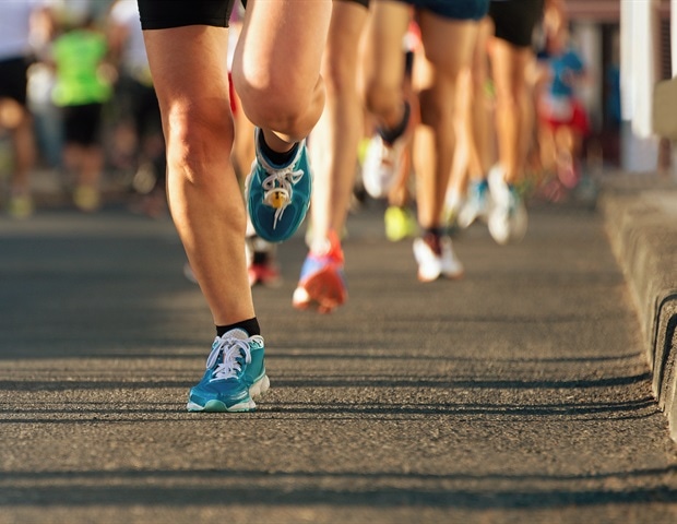
  • How marathon running in novices affects the blood vessels.
  • Machine learning to diagnose pulmonary arterial hypertension.
  • Feasibility of free-breathing MRI without sedation in newborns.
  • CMR for more sensitive assessment of cardiotoxicity of cancer drugs.


Explore the scientific programme to discover innovative studies on how CMR – also called cardiac magnetic resonance imaging (MRI) – is being used to fine tune diagnosis and treatment of cardiovascular disease.


Five late-breaking studies will be presented on the use of CMR to improve risk prediction and treatment of patients with heart disease:


  • DREAM trial on reducing heart damage and improving recovery in heart attack patients who have undergone successful treatment with percutaneous coronary intervention (PCI).
  • T-TIME trial on use of a clot-busting drug during PCI in heart attack patients to reduce blockages in small blood vessels.
  • The large Hypertrophic Cardiomyopathy Registry in six countries using CMR, genetics, and biomarkers to improve prediction of risk for sudden cardiac death and heart failure.
  • Can assessment of myocardial fibrosis (scarring of the heart muscle) using CMR predict outcomes in patients who have undergone heart transplantation?
  • Can patients with legacy pacemaker and defibrillator leads safely undergo MRI?


“All of these studies use CMR to tackle important clinical questions,” said Dr José Rodriguez-Palomares, Programme Chair. “The DREAM and T-TIME trials attempt to improve outcomes after a heart attack by combining opening of the blocked artery (PCI) with a drug or intervention. CMR may be a new way to risk stratify patients with hypertrophic cardiomyopathy and the registry will provide insights into this emerging area. Heart transplant recipients are intensely monitored for rejection and CMR may be a non-invasive mode of assessment. The final study addresses concerns about MRI interfering with cardiac devices.”


EuroCMR will attract more than 1,000 cardiologists, radiologists, radiographers, imaging scientists, MRI physicists, computer scientists and engineers from over 65 countries. Global experts will present the latest research across 2.5 days and 65 sessions.


The conference theme is “Extending the clinical value of CMR through quality and evidence”. Dr Rodriguez-Palomares said: “In the past CMR was a complementary technique to echocardiography to optimise diagnosis in patients with cardiac diseases. Now, on top of that, it is increasingly used to risk stratify patients, tailor treatment, and improve prognosis.”


C-Path and FARA announce launch of Friedreich’s Ataxia Integrated Clinical Database

A banner of news medical and life science

Critical Path Institute’s (C-Path) Data Collaboration Center (DCC) and the Friedreich’s Ataxia Research Alliance (FARA) today announced the launch of the Friedreich’s Ataxia Integrated Clinical Database (FAICD). The new platform will enable collaborative research and data sharing to support the understanding of natural history, potential biomarkers and clinical endpoints, and promote research into novel clinical trial design in Friedreich’s ataxia (FA). By making this data available to researchers, the organizations hope to enable the development of tools that will help design and interpret efficient clinical trials — leading to effective treatments for FA as soon as possible.


“FA is a rare, progressive and fatal disease that affects multiple organ systems, and those living with the disease are in urgent need of effective treatments,” explained FARA’s Executive Director Jennifer Farmer. “FAICD addresses this need by providing a platform to share data and making it available to qualified researchers to expedite the drug development process. We believe this resource will inform and give future clinical trials of potential therapies the best chance of success.”


FAICD contains data contributed by collaborating companies that have carried out clinical trials in FA, as well as natural history and clinical outcome measure data from the Friedreich’s Ataxia Clinical Outcomes Measures Study (FACOMS) collected by the Collaborative Clinical Research Network in FA and funded by FARA.


All data contributed to this project is de-identified, mapped to standards defined by the Clinical Data Interchange Standards Consortium (CDISC) and curated by C-Path’s DCC before inclusion in FAICD.


“C-Path has a long history of expertise in data standards development, curation, and oversight of multiple data sharing initiatives,” said Richard Liwski, Director of the DCC and C-Path’s Chief Technology Officer. “We are proud to be part of the effort to coordinate collaborative contributions from data owners and integrate that data into a single database for this rare, degenerative and life-shortening neuromuscular disorder.”


Study unravels genetic basis of hypertrophic cardiomyopathy cases in Finland

Study unravels hypertrophic cardiomyopathy cases in Finland

One third of hypertrophic cardiomyopathy cases in Finland are caused by one of the four major mutations, a new study from the University of Eastern Finland and Kuopio University Hospital shows. Overall, 40% of patients carried a specific or a likely mutation causing the disease, and 20% were carriers of a rare gene mutation whose role in the disease remains unknown. The findings of the nationwide FinHCM study, led by Professor Johanna Kuusisto at the University of Eastern Finland, were published in ESC Heart Failure.


The study is the most extensive one so far to analyze the genetic background and outcome of hypertrophic cardiomyopathy in a nationwide cohort.


Hypertophic cardiomyopathy is estimated to occur in one in 500 adults. Among young people and athletes, it is the most common cause of sudden cardiac deaths. According to the newly published study, the prognosis of patients diagnosed with and treated for hypertrophic cardiomyopathy in Finland is good. In a seven-year follow-up, their mortality rate was low, although significantly higher than the mortality rate of the age- and sex-controlled population.


The researchers also analyzed the National Cause of Death Register to explore the role of hypertrophic cardiomyopathy in all deaths occurring in Finland. Over a ten-year period, almost 600 Finns died of hypertrophic cardiomyopathy, and one third of these deaths were sudden cardiac deaths.


According to Professor Kuusisto, the high prevalence of sudden cardiac deaths indicates that cardiomyopathy remains underdiagnosed. “In order to prevent sudden cardiac deaths in particular, it is vital that patients get a timely diagnosis and proper treatment,” she says.


The researchers studied gene mutations associated with hypertrophic cardiomyopathy in a nationwide cohort of 382 people by using targeted sequencing and a gene panel comprising a total of 59 genes associated with the disease. The outcome of the disease was analyzed in a follow-up study involving 428 participants.


The genetic background of the disease can differ from one population to the next. Professor Kuusisto’s research group has studied the genetics, clinical picture and pathogenesis of hypertrophic cardiomyopathy (HCM) and dilated cardiomyopathy (DCM) in Finland since the 1990s. The four Finnish major mutations causing hypertrophic cardiomyopathy have been discovered in the nationwide FinHCM study led by Professor Kuusisto.


Research has also advanced the diagnostics and treatment of cardiomyopathies. “The use of gene diagnostics, magnetic resonance imaging of the heart and implantable cardioverter-defibrillators has improved the outcome of patients,” Professor Kuusisto says.


The research group’s new projects involve induced pluripotent stem cell (iPS) technology, among other things. The researchers use cardiomyocytes developed with iPS to analyze cellular-level effects in the major mutations, and reasons behind different clinical presentations in the carriers of the same gene mutation.


Ultromics expands multiple clinical trials for coronary heart disease to the U.S.

An artificial heart kept on a doctors report

Ultromics, the U.K. start-up behind the world’s first outcomes driven, AI-based, ultrasound diagnostic support tool for coronary artery disease (CAD), is currently supporting multiple ongoing clinical trials.

These trials include “Rainier”, based in the U.S., and “EVAREST”, which is expanding across 35 NHS sites within the U.K.

Both trials focus on the diagnosis of coronary artery disease using Ultromics’ first product, EchoGo. Results are expected within the next few months and will be submitted to the FDA with the intention of obtaining clearance for sale within the U.S.

The U.S. based Rainier trial includes a retrospective review of 550 stress exams, as well as a total of 1,600 cardiologist reads from a variety of skill levels.

The first phase of the trial is located at Oregon Health and Science University (OHSU) and is being accomplished in conjunction with leading cardiologists Dr. Sanjiv Kaul, Director of the Knight Cardiovascular Institute, and Dr. Stephen Heitner, Director of the Hypertrophic Cardiomyopathy Clinic.

The U.K. based EVAREST trial – administered by the Oxford Cardiovascular Clinical Research Facility at the University of Oxford – has successfully entered Phase III.

Phase III will deliver recruitment of 5,000 participants across 35 NHS sites, making it one of the largest echo studies in the U.K.

This phase sees continued recruitment from Phase II, with 11 new sites and a further 15 sites currently undergoing feasibility assessment to join in early 2019. This will allow for the generalisability of stress echo protocols, machine types, operators, and patient groups across different healthcare settings.

At the conclusion of these trials, Ultromics intends to prove that EchoGo is the world’s most accurate echocardiography-based tool for the diagnosis of CAD.

Traditionally, clinicians use echocardiograms to diagnose a patient’s condition based on just a few factors, which leads to an accurate diagnosis roughly 80% of the time under the best circumstances. EchoGo, on the other hand, uses deep learning and one of the world’s largest echo image databases.

It supports clinicians by delivering greater accuracy in both sensitivity and specificity and could save healthcare payers and providers significant amounts by reducing the incidence of patients undergoing unnecessary surgical procedures.

Just as significantly, it can save lives by preventing patients with potentially fatal heart disease from being sent home.


Heart disease is the biggest killer globally. Of the 3 million stress exams performed in the US each year, it is estimated around 600,000 may be misread.

This is potentially costing the US healthcare industry billions of dollars in unnecessary additional costs.

We are determined to provide patients and providers with a reliable diagnosis, which in turn will create significant improvement to patient outcomes and savings for healthcare systems.”

Ross Upton, CEO and Founder at Ultromics


Researchers discover why heart contractions are weaker in individuals with HCM

A human heart with all its veins with a white background

When a young athlete suddenly dies of a heart attack, chances are high that they suffer from familial hypertrophic cardiomyopathy (HCM). Itis the most common genetic heart disease in the US and affects an estimated 1 in 500 people around the world. A protein called myosin acts as the molecular motor which makes the muscles in the heart contract. Researchers had suspected for some time that the R403Q mutation in some of the myosin genes is among those that play a role in causing HCM. But experiments using mice models failed to show that this was indeed the case. (Mice are often used in experiments because their behavior, biology and genetic material resemble those of humans).

An international team led by Professor Dilson Rassier from McGill’s Department of Kinesiology and Physical Education, has discovered, by working with transgenic rabbits with the R403Q mutation, that in these rabbits, individual myosin molecules and myofibrils (the basic rod-like filaments inside muscles) produce less force and a lower maximum velocity of contraction than those isolated from healthy hearts.

They reached this conclusion by using advanced techniques such as atomic force microscopy and molecular motility assays (which allows them to visualize the movements of these proteins in vitro) to look more closely at what was going on within myosin molecules and also within myofibrils.

“It’s been difficult to gain a clear picture of what is going on within the myosin proteins with this mutation, simply because of the technical and experimental limitations of looking closely at objects of this minute size and measuring their force and motility (myosin molecules are about 19 nanometers (0.0000019 centimeters),” explains Rassier. “The results should help clinicians develop drugs and chemicals that target this specific function of myosin in future.”

https://www.mcgill.ca/newsroom/channels/news/why-heart-contractions-are-weaker-those-hypertrophic-cardiomyopathy-290647


New rapid imaging protocol quickly and cheaply detects heart ailments for people in Peru

New imaging protocol detects heart ailments for people

A newly developed rapid imaging protocol quickly and cheaply diagnosed heart ailments in patients in Peru, according to new research in Journal of the American Heart Association, the Open Access Journal of the American Heart Association/American Stroke Association.

In Peru, cardiovascular disease affects 3.2 million (16 percent of the adult population), leading to a significant loss of well-being, estimated at 281,829 Disability Adjusted Life Years.

Current Cardiac Magnetic Resonance Imaging (CMR) usage in high income countries focuses on function, scar (fibrous tissue that replaces normal heart muscle tissue destroyed by injury or disease), perfusion, flow, mapping and angiography and delivers valuable clinical insights leading to targeted and precise treatments. However, these varied techniques make it slow (typically 45 minutes), expensive, complex and potentially out of reach for most people in the developing world.

In this study, researchers developed and tested a rapid CMR protocol using contrast dye that measured cardiac structure, function and scarring. The rapid diagnostics worked with existing infrastructure, took 18 minutes and cost $150 per patient resulting in important changes in patient care.

“Our CMR strategy was three to five times cheaper than current CMR exams in Peru,” said James C. Moon, M.D., study lead author and professor at Barts Heart Centre, St. Bartholomew’s Hospital in London. “It also can be delivered two to three times faster and is easier than conventional CMR.”

Researchers conducted scans on 98 Peruvian patients (average age 52, 60 percent female). Scanning found 26 percent had hypertrophic cardiomyopathy, 22 percent had dilated cardiomyopathy, 15 percent had ischemic cardiomyopathy (when cardiovascular disease or heart attack cause the disease), and uncovered 12 other pathologies including tumors, congenital heart disease, iron overload, amyloid plaques (abnormal protein deposits), genetic syndromes, inflamed vessels, clots and valve disease.

Researchers report CMR revealed an unsuspected new diagnosis in 19 percent of patients or led to a change of treatment in 37 percent. In 5 percent, a change in care management was suggested but not delivered due to access barriers (cardiac surgery or device therapy).

Researchers found rapid CMR satisfied all imaging needs in 89 percent of patients. In 7 percent where CMR was the first imaging technique performed, no further non-invasive imaging was needed. CMR did not miss any diagnoses initially found by echocardiography, researchers said.

“Because the rapid CMR protocol was embedded in clinical care with training and education, it resulted in important and frequent patient management changes that appeared beneficial for both patients and the healthcare system,” said Katia Menacho, Ph.D., study first author and cardiovascular science research fellow at Barts Heart Centre, St. Bartholomew’s Hospital in London. “Lack of resources is not a justification for the absence of key diagnostic tests in the developing world.”

In an accompanying editorial, Christopher M. Kramer, M.D., University of Virginia Health System in Charlottesville notes: “To make this proof-of-principle study a reality in much of the developing world, imagers will need to be trained at sites with appropriate scanner technology. Only in this way will an abbreviated protocol for evaluation of cardiomyopathies be implemented. This is an exciting time for the potential of broadening the impact of CMR throughout the developing world.”

https://newsroom.heart.org/news/rapid-heart-imaging-technique-may-cut-costs-boost-care-in-developing-world?preview=ee26


Seattle Children’s opens new clinic to provide convenient access to pediatric specialty care services

Four children jumping with a sunset behind

Seattle Children’s today announced the opening of its newest regional clinic, Seattle Children’s North Clinic, which will provide convenient access to pediatric specialty care services for families in north King, Snohomish, Whatcom and Skagit counties. The 37,000-square-foot clinic is located on Providence Regional Medical Center Everett’s Colby Campus at 1815 13th St.

The new clinic will provide services in more than 18 pediatric specialties, including audiology, otolaryngology, cardiology, rehabilitation and sports therapy, among others. It will also include an urgent care clinic, offering same-day appointments, including holidays, an imaging center, Child Wellness Clinic, and playroom for patients and siblings.

“We’re committed to meeting the needs of families in the north by providing world-class care closer to home,” said David Stolte, director of North Sound Regional Clinics. “Families will no longer have to choose between getting pediatric-focused care and convenience. We’ll be located where patients and families need us, in their community.”

Seattle Children’s North replaces both Seattle Children’s Everett and Mill Creek clinics, which closed as of Aug. 10, adding ten new pediatric services to the North Sound area. In addition to Seattle Children’s North, Seattle Children’s has specialty regional clinics in Bellevue, Federal Way, Olympia, Tri-Cities and Wenatchee.

Kara Chitwood said Seattle Children’s will always hold a special place in her heart. When her son, Bretton, broke his ankle in nine places in 2016 he was rushed to Seattle Children’s Mill Creek Clinic from their home in Lynden, Washington. He needed to be closely monitored after his initial visit, and so the family found themselves traveling to the clinic nearly every week for appointments.

“We’ve always had wonderful experiences at Seattle Children’s,” said Chitwood. “We wouldn’t go anywhere else.”

Chitwood credits the expert care they received at the clinic for saving her son’s life. After returning to play hockey after his ankle healed, Bretton experienced lingering pain. Chitwood called their provider at the clinic who ordered imaging for the next day. Later that week, the family found out Bretton had osteosarcoma, an aggressive bone cancer.

“He could have had a very different outcome if we wouldn’t have caught it so early,” said Chitwood.

Bretton recently graduated high school and is preparing for college in the fall.

“As parents, we want to make sure our children receive the best care,” said Chitwood. “Knowing how well Seattle Children’s ranks both regionally and nationally, it’s where we’ll continue to go. I could take my children somewhere else, or I could go to Seattle Children’s North Clinic where I know we’ll get better care and faster.”

Jennifer Campbell’s 6-year-old daughter, Hannah, has been a patient at Seattle Children’s since birth. Only 12 hours after being born, Hannah was transferred to Seattle Children’s with a rapid heartbeat and diagnosed with supraventricular tachycardia (SVT), a rapid heartbeat that can be common in newborns, and hypertrophic cardiomyopathy, a thickening of the heart muscle. After a couple weeks, Hannah was sent home for the first time, but her condition worsened. Her heart doubled in size and was failing. She need needed a heart transplant and quickly. Doctors weren’t sure she’d make it to transplant, but on Sept. 23, 2012, they got the call. At only 4 months old, Hannah received a new heart.

“Seattle Children’s saved our daughter’s life,” said Campbell.

Today, Hannah is 6 years old and thriving. Campbell said she is excited for Seattle Children’s North to open. The clinic is only a 10-minute drive from their home.

Hannah needs to get her blood drawn nearly every month, and so the lab at the clinic will be much more convenient for the family, rather than having to drive to Seattle Children’s main campus.

“We’re going to be at the clinic a lot,” said Campbell. “From fevers to blood draws, it will be nice to have a place we’re familiar with and trust so close to home. Seattle Children’s really is like an extension of our family.”

http://www.seattlechildrens.org/


Combining stem cell technology and artificial intelligence to diagnose genetic cardiac diseases

Combining stem cell technology and artificial intelligence

A new study by Professors Martti Juhola and Katriina Aalto-Setälä of the University of Tampere in Finland demonstrates that with the use of artificial intelligence and machine learning, it is possible not only to accurately sort sick cardiac cell cultures from healthy ones, but also to differentiate between genetic cardiac diseases.

iPSC-derived cardiomyocytes can be derived from a blood sample or a skin biopsy. These cells are currently used to understand the pathophysiology of different diseases and to identify new potential drugs for various diseases.

Machine learning and artificial intelligence have greatly improved in recent years. Scientists at the University of Tampere have now combined stem cell technology and artificial intelligence to study beating cardiomyocytes in cell cultures. The beating behavior of the cells was analyzed using calcium signals. Calcium is essential for cardiomyocytes to beat, and the beating can be monitored by using fluorescent labels.

In the study, the cardiomyocytes were derived either from patients with a genetic arrhythmia (CPVT), long QT syndrome (LQTS), or hypertrophic cardiomyopathy (HCM), or from healthy individuals. The beatings of single cardiomyocytes were recorded and the analysis software was taught what diseases they represented. The program then learned to separate the different groups and to identify specific features in the beating behavior of each cell.

The software is now capable of identifying whether signals are from cells derived from an individual carrying a disease-causing mutation or from a healthy individual. This is very impressive, but the biggest surprise was that the program could also tell the difference between the diseases.

This important observation reveals that iPSC-derived cells and artificial intelligence have the potential to be used in diagnostics. Currently, genetic diseases are mainly diagnosed by DNA analysis, but in many cases the results do not reveal whether the DNA alteration is the true cause of the disease or whether it is just an innocent variation. This new finding demonstrates that uniting artificial intelligence and machine learning can help in such situations. The combination of technologies could also be used in cases of unspecific but severe cardiac findings to identify the specific disease causing the symptoms.

http://www2.uta.fi/en/news/story/diagnostics-genetic-cardiac-diseases-using-stem-cell-derived-cardiomyocytes


Study combines gene editing and stem cell technologies to predict person’s risk for heart disease

An artificial heart kept on a report

Scientists may now be able to predict whether carrying a specific genetic variant increases a person’s risk for disease using gene editing and stem cell technologies, according to new research in the American Heart Association’s journal Circulation.

For the first time, the study demonstrates the unique potential of combining stem cell-based disease modeling (Induced pluripotent stem cells) and CRISPR/Cas9-mediated genome editing technology as a personalized risk-assessment platform for determining the disease-causing ability of a yet undescribed genetic variant, known as a “variant of uncertain significance” or VUS.

Numerous genetic variations are identified as “related” to a medical condition, but it is uncertain if they actually lead to disease, said study senior author Joseph C. Wu, M.D., Ph.D., director of the Stanford Cardiovascular Institute and Simon H. Stertzer, M.D. Endowed Professor in the Department of Medicine (Cardiology) and Department of Radiology at the Stanford University School of Medicine in California.

“Random genetic testing will create a lot of stress for a healthy individual who may be getting echocardiograms, MRIs or medications that they may not need,” Wu said. “Results from this study will help improve the interpretation and diagnostic accuracy of gene variants, especially in the era of personalized medicine and precision health. The goal is to optimize the decision making of clinicians in their choices of therapy by providing a much clearer result for the ‘variant of uncertain significance’ carriers.”

Researchers studied genetic variants associated with hypertrophic cardiomyopathy, a condition in which the heart muscle thickens. It is a common cause of sudden cardiac death in young people and young athletes.

They harvested DNA from 54 “healthy” or symptom-free individuals without heart disease, then sequenced their DNA using a custom DNA panel of 135 cardiomyopathy and congenital heart disease genes associated with sudden cardiac death.

The sequence results uncovered 592 unique genetic variants, with 78 percent of genetic variants being classified as “benign,” “likely benign,” or a “variant of uncertain significance.” However, 17 genetic variants were annotated as “likely pathogenic” or disease-causing.

One individual who had multigenerational family members carrying a variation in gene MYL3, which is associated with hypertrophic cardiomyopathy, was chosen in this study.

After collecting the patients’ peripheral blood mononuclear cells, the cells were reprogrammed to induced pluripotent stem cells (iPSCs) and genome edited using the CRISPR/Cas9 gene editing technology to engineer cells with the same genetics (isogenic iPSC lines). Comprehensive analysis was next performed on the engineered cell lines to determine the MYL3 variant could lead to disease.

Traditionally, treating hypertrophic cardiomyopathy depends on whether a patient has symptoms and the severity of those symptoms. People who have “silent” hypertrophic cardiomyopathy without symptoms are not treated. For those with symptoms, physicians may recommend lifestyle changes such as adopting a heart-healthy diet, reducing stress and incorporating exercise while treating underlying illnesses that can make the condition worse. They may also prescribe medications for hypertrophic cardiomyopathy, typically reserving surgery for more severe cases.

“Given the diversity of the human genome – no one of us is identical to another – it is difficult to determine whether a genetic “variant” is meaningful or not. As a result, we risk treating patients with medications or more for a variant that, in the end, is benign,” said Circulation editor, Joseph A. Hill, M.D., Ph.D., chief of cardiology at UT Southwestern Medical Center in Dallas. “This study combined two new powerful technologies, induced pluripotent stem cells and CRISPR-Cas9 gene editing, to model a patient’s heart in a dish and to test whether those heart cells manifested signs of disease. This approach heralds a new era of in vitro disease modeling and drug testing as pivotal elements of precision medicine.”

https://newsroom.heart.org/news/gene-editing-technology-may-improve-accuracy-of-predicting-individuals-heart-disease-risk?preview=d22c


Gene editing technology predicts heart disease risk

Gene editing technology predicts heart disease risk

Scientists may now be able to use gene editing and stem cell technologies to predict whether carrying a specific gene variant increases a person’s risk of heart disease, according to a new study published in the American Heart Association’s journal Circulation.

This is the first time a study has demonstrated the potential of combining stem cell-based disease modeling with CRISPR/Cas 9-mediated genome editing, to determine the pathogenic ability of an undescribed genetic variant known of as “variant of uncertain significance” (VUS).

Many genetic variants are known to be related to medical conditions, but whether they actually lead to disease remains unknown.

Senior author Joseph Wu (Stanford University School of Medicine, California) says random genetic testing creates a lot of stress for healthy individuals who may be getting echocardiograms, MRIs or medications that they may not need.


The goal is to optimize the decision making of clinicians in their choices of therapy by providing a much clearer result for the ‘variant of uncertain significance’ carriers.”

Joseph Wu, Senior Author


For the study, the researchers looked at genetic variants associated with hypertrophic cardiomyopathy; a common cause of sudden death in young people and athletes.

They took DNA from 54 symptom-free individuals without heart disease and sequenced it using a panel of 135 cardiomyopathy and congenital heart disease genes associated with sudden cardiac death.

The results revealed 592 unique genetic variants, of which 78% were classified as benign, likely benign or a VUS. However, 17 variants were classified as likely pathogenic.

One participant had family members who were carrying a variant in the gene MYL3, which is associated with hypertrophic cardiomyopathy.

The researchers collected that patient’s blood mononuclear cells and reprogrammed them to induce pluripotent stem cells.

They then edited the genome of the cells using CRISPR/Cas9 gene editing to engineer cells with the same genetics. Those engineered cells could then be analysed to determine whether MYL3 could cause disease.


This study combined two new powerful technologies, induced pluripotent stem cells and CRISPR-Cas9 gene editing, to model a patient’s heart in a dish and to test whether those heart cells manifested signs of disease. This approach heralds a new era of in vitro disease modeling and drug testing as pivotal elements of precision medicine.”

Joseph Hill, Editor of Circulation


https://www.eurekalert.org/pub_releases/2018-06/aha-get061318.php