New book focuses on heart failure in pediatric patients

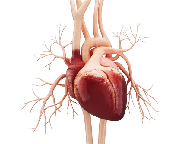
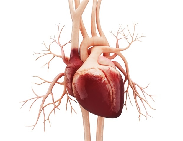
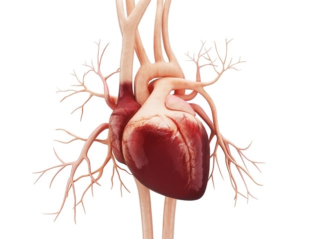
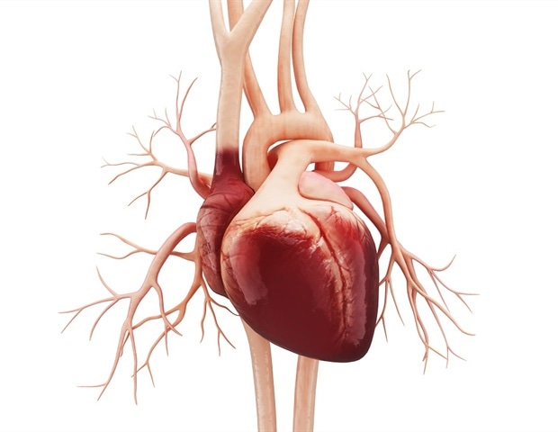
A human heart with all its veins with a white background

The field of heart failure in pediatric populations has recently initiated more registries and clinical trials. This age group has special concerns and particularities regarding their clinical picture and management. Therefore, multiple disciplines have been involved in the management of heart failure in this age group.

The book Heart Failure in Pediatric Patients will solve various issues faced by professionals dealing with infants and children with cardiac problems. The cases discussed are supplemented with videos and images providing close proximity to real time experience.

Pathophysiology, classification and clinical presentation focusing on congenital heart diseases has been discussed providing different modes of clinical investigations including heart failure in conditions of hypertrophic cardiomyopathy.

This new volume in the book series on heart failure medicine also includes detailed radiological and imaging investigations for heart failure in general; however, it also discusses the investigation of certain pediatric and congenital heart disease. Cardiology learners at different levels; undergraduate, postgraduate, specialists and allied professionals will gain substantial knowledge obtained from the perspective of numerous cardiologists practicing in different regions.

Study traces the molecular roots of hypertrophic cardiomyopathy

A human heart with all its veins with a white background

The heart’s ability to beat normally over a lifetime is predicated on the synchronized work of proteins embedded in the cells of the heart muscle.

Like a fleet of molecular motors that get turned on and off, these proteins cause the heart cells to contract, then force them to relax, beat after life-sustaining beat.

Now a study led by researchers at Harvard Medical School, Brigham and Women’s Hospital and the University of Oxford shows that when too many of the heart’s molecular motor units get switched on and too few remain off, the heart muscle begins to contract excessively and fails to relax normally, leading to its gradual overexertion, thickening and failure.

Results of the work, published Jan. 27 in Circulation, reveal that this balancing act is an evolutionary mechanism conserved across species to regulate heart muscle contraction by controlling the activity of a protein called myosin, the main contractile protein of the heart muscle.

The findings- based on experiments with human, mouse and squirrel heart cells- also demonstrate that when this mechanism goes awry it sets off a molecular cascade that leads to cardiac muscle over-exertion and culminates in the development of hypertrophic cardiomyopathy (HCM), the most common genetic disease of the heart and a leading cause of sudden cardiac death in young people and athletes.

Our findings offer a unifying explanation for the heart muscle pathology seen in hypertrophic cardiomyopathy that leads to heart muscle dysfunction and, eventually, causes the most common clinical manifestations of the condition.”

Christine Seidman, senior author, professor of genetics in the Blavatnik Institute at Harvard Medical School, a cardiologist at Brigham and Women’s Hospital and a Howard Hughes Medical Institute Investigator

Importantly, the experiments showed that treatment with an experimental small-molecule drug restored the balance of myosin arrangements and normalized the contraction and relaxation of both human and mouse cardiac cells that carried the two most common gene mutations responsible for nearly half of all HCM cases worldwide.

If confirmed in further experiments, the results can inform the design of therapies that halt disease progression and prevent complications.

“Correcting the underlying molecular defect and normalizing the function of heart muscle cells could transform treatment options, which are currently limited to alleviating symptoms and preventing worst-case scenarios such as life-threatening rhythm disturbances and heart failure,” said study first author Christopher Toepfer, who performed the work as a postdoctoral researcher in Seidman’s lab and is now a joint fellow in the Radcliffe Department of Medicine at the University of Oxford.

Some of the current therapies used for HCM include medications to relieve symptoms, surgery to shave the enlarged heart muscle or the implantation of cardioverter defibrillators that shock the heart back into rhythm if its electrical activity ceases or goes haywire. None of these therapies address the underlying cause of the disease.

Imbalance in the motor fleet

Myosin initiates contraction by cross-linking with other proteins to propel the cell into motion. In the current study, the researchers traced the epicenter of mischief down to an imbalance in the ratio of myosin molecule arrangements inside heart cells. Cells containing HCM mutations had too many molecules ready to spring into action and too few myosin molecules idling standby, resulting in stronger contractions and poor relaxation of the cells.

An earlier study by the same team found that under normal conditions, the ratio between “on” and “off” myosin molecules in mouse heart cells is around 2-to-3. However, the new study shows that this ratio is off balance in heart cells that harbor HCM mutations, with disproportionately more molecules in active versus inactive states.

In an initial set of experiments, the investigators analyzed heart cells obtained from a breed of hibernating squirrel as a model to reflect extremes in physiologic demands during normal activity and hibernation. Cells obtained from squirrels in hibernation-;when their heart rate slows down to about six beats per minute-;contained 10 percent more “off” myosin molecules than the heart cells of active squirrels, whose heart rate averages 340 beats per minute.

“We believe this is one example of nature’s elegant way of conserving cardiac muscle energy in mammals during dormancy and periods of deficient resources,” Toepfer said.

Next, researchers looked at cardiac muscle cells from mice harboring the two most common gene defects seen in HCM. As expected, these cells had altered ratios of “on” and “off” myosin reserves. The researchers also analyzed myosin ratios in two types of human heart cells: Stem cell-derived human heart cells engineered in the lab to carry HCM mutations and cells obtained from the excised cardiac muscle tissue of patients with HCM. Both had out-of-balance ratios in their active and inactive myosin molecules.

Further experiments showed that this imbalance perturbed the cells’ normal contraction and relaxation cycle. Cells harboring HCM mutations contained too many “on” myosin molecules and contracted more forcefully but relaxed poorly. In the process, the study showed, these cells gobbled up excessive amounts of ATP, the cellular fuel that sustains the work of each cell in our body. And because oxygen is necessary for ATP production, the mutated cells also devoured more oxygen than normal cells, the study showed. To sustain their energy demands, these cells turned to breaking down sugar molecules and fatty acids, which is a sign of altered metabolism, the researchers said.

“Taken together, our findings map out the molecular mechanisms that give rise to the cardinal features of the disease,” Seidman said. “They can help explain how chronically overexerted heart cells with high energy consumption in a state of metabolic stress can, over time, lead to a thickened heart muscle that contracts and relaxes abnormally and eventually becomes prone to arrhythmias, dysfunction and failure.”

Restoring balance

Treating both mouse and human heart cells with an experimental small-molecule drug restored the myosin ratios to levels comparable to those in heart cells free of HCM mutations. The treatment also normalized contraction and relaxation of the cells and lowered oxygen consumption to normal levels.

The drug, currently in human trials, restored myosin ratios even in tissue obtained from the hearts of patients with HCM. The compound is being developed by a biotech company; two of the company’s co-founders are authors on the study. The company provided research support for the study.

In a final step, the researchers looked at patient outcomes obtained from a database containing medical information and clinical histories of people diagnosed with HCM caused by various gene mutations. Comparing their molecular findings from the laboratory against patient outcomes, the scientists observed that the presence of genetic variants that distorted myosin ratios in heart cells also predicted the severity of symptoms and likelihood of poor outcomes, such as arrhythmias and heart failure, among the subset of people that carried these very genetic variants.

What this means, the researchers said, is that clinicians who identify patients harboring gene variants that disrupt normal myosin arrangements in their heart muscle could better predict these patients’ risk of adverse clinical course.

“This information can help physicians stratify risk and tailor follow-ups and treatment accordingly,” Seidman said.

New program receives national recognition for treating hypertrophic cardiomyopathy

recognition for treating hypertrophic cardiomyopathy

When Kayla Green was first diagnosed with hypertrophic cardiomyopathy (HCM), she felt vindicated.

“For years, I could feel something was wrong in my chest,” she said. “Other people insisted that I was just having panic attacks, but I knew it was something else. No one was taking me seriously, and it wasn’t until after a trip to the emergency room during college that I finally had an electrocardiogram (EKG) and I was diagnosed with HCM.”

Her journey with HCM has had its ups and downs, but thanks to the high quality of care she’s received at UC San Diego Health, she says she finally feels like her treatment is being taken seriously.

The care received by Green and patients like her is why the Hypertrophic Cardiomyopathy Association (HCMA) is recognizing UC San Diego Health this week as a Center of Excellence in treating the condition, one of only four such centers in the state. The HCMA is an international resource for patients, families and the medical community. The organization provides services to enhance understanding, foster research and ensure high quality health care to HCMA patients. The HCMA Center of Excellence recognition acknowledges medical centers that offer patients with the condition the most comprehensive care.

We are honored to receive this recognition. We are dedicated to providing comprehensive care for HCM patients like Kayla and hope that this recognition will raise awareness in the San Diego community about the availability of HCM screening and treatment at UC San Diego Health.”

Jorge Silva Enciso, MD, cardiologist at the Cardiovascular Institute at UC San Diego Health

Green is not alone in her experience. HCM affects approximately one in every 250 people. The condition is caused by abnormal thickening of the muscle in the heart, which makes it harder for the heart to pump blood throughout the body. Often, HCM goes undiagnosed because for many people, there are no obvious symptoms -; or the symptoms are attributed to other conditions.

When symptoms are present, they can be severe and life-threatening. HCM can lead to chest pain, shortness of breath, and most critically, abnormal heart rhythms known as arrhythmias. For some, the first warning sign is having a close relative die unexpectedly in their sleep; the condition is highly heritable, with an approximately 50 percent chance of a parent passing it on to their child.

With leadership by Silva Enciso and Eric Adler, MD, director of cardiac transplant and mechanical circulatory support, the UC San Diego Health hypertrophic cardiomyopathy program offers a wide range of diagnostic and treatment options, with a variety of experts, including cardiologists, surgeons, genetic counselors and mental health professionals all aimed at supporting HCM patient care.

“Our heart transplant program is the largest in San Diego, and our position as an academic medical center means that there are researchers right on campus working at better understanding this condition and developing new clinical trials so we can provide better care to patients in the future,” said Silva Enciso.

“The Hypertrophic Cardiomyopathy Association is pleased to welcome UC San Diego Health to our family of Center of Excellence programs,” said Lisa Salberg, founder and CEO of HCMA. “The HCMA has worked hard over the past 24 years to ensure that HCM patients receive the highest quality care in high volume programs. Patients and families benefit by having a team of highly skilled clinicians, as well as the emotional support they need to manage living with a genetic heart condition that may impact many other members of the family as well.”

For patients like Green, that kind of support has been critical to her recovery. “Before I came to UC San Diego Health, I was only seeing a cardiologist once a year and taking some blood pressure medications,” she said. “Doctors never did any screening beyond an EKG to see how severe my HCM was, or gave me much information about long-term care.

“I have really liked working with Dr. Silva Enciso and his team. He’s the first doctor in my whole life to help me understand my condition. He showed me what my actual heart looks like and explained what this condition means -; and the additional screenings he recommended helped us see how the HCM has spread and figure out next steps for my treatment.”

As an HCMA Center of Excellence, UC San Diego Health will continue to provide comprehensive services for HCM patients within the San Diego community. Green hopes that more patients will take advantage of HCM screening and treatment resources.

“Being diagnosed as a young person, you realize how serious this condition is,” she said. “It’s so underdiagnosed, and people die from it all the time without anyone ever realizing. I really think that there should be routine screening for HCM, like there is for cancer. You just never know if you’re at risk.”

Researchers reveal initial results from a study of hypertrophic cardiomyopathy

A doctor standing writing on a report

Researchers have revealed the initial results from the world’s largest comprehensive study of hypertrophic cardiomyopathy, an abnormal thickening of the heart that often goes undiagnosed and can prove deadly. The condition can present at any age, and it is the most common cause of sudden cardiac death in young athletes.

Researchers reveal initial results from a study of hypertrophic cardiomyopathy

UVA’s Christopher M. Kramer, MD, is a co-principal investigator of the massive study of hypertrophic cardiomyopathy.

The early findings from the $14.4 million study suggest that patients can largely be grouped into two buckets: Patients with a clearly defined genetic mutation tended to have more scarring of the heart muscle, while patients without such a mutation tended to have no scar and more obstruction of blood flow. That information improves doctors’ understanding of the condition and will, with additional research, help them predict patients’ risk of sudden cardiac death and heart failure and determine the best treatment strategies.

It really changes the way we think about patients. We can categorize them more easily. The more we can understand and group patients into categories, the better we will be able to learn what the best therapies are.”

Christopher M. Kramer, MD, a cardiologist at UVA Health and a co-principal investigator of the massive study

Bold new approach

The researchers developed and examined a registry of more than 2,750 patients with hypertrophic cardiomyopathy (HCM) at 44 sites in six countries. In a first-of-its-kind approach, the research team integrated high-tech imaging, genetic analysis and biomarker data with traditional clinical information to facilitate the most sophisticated analysis of HCM ever undertaken.

Most prior HCM analyses have been retrospective, meaning they examined pre-existing data. That approach is limited by what information happens to be available. In contrast, the creators of the new registry – officially known as the Hypertrophic Cardiomyopathy Registry – have thoughtfully collected a wide spectrum of useful data from the study participants so that researchers can finally see the bigger picture.

The project has already generated a gold mine of information, and the research team members expect the data will require years of analysis. But the end result should have concrete benefits for patients: The researchers believe that the registry will ultimately let them identify key clues to determine which patients are at greatest risk and which treatments will best benefit different groups of patients.

Take scarring in the heart, for example: “We think scar, which we can identify with MRI of the heart, is a powerful predictor of bad outcomes,” Kramer said. But, he noted, scarring is just one factor, and the registry will let doctors look at hypertrophic cardiomyopathy’s myriad complexities in a much more sophisticated and nuanced manner.

We look at this data and everything is making more sense. It’s all fitting together.”

Christopher M. Kramer, MD

Findings published

The researchers have published their early findings in the Journal of the American College of Cardiology.

Journal reference:

Neubauer, S. et al. (2019) Distinct Subgroups in Hypertrophic Cardiomyopathy in the NHLBI HCM Registry. Journal of the American College of Cardiology. doi.org/10.1016/j.pubrev.2019.101835

Study finds no overall improvement in heart transplant waitlist after policy change

A human heart with all its veins with a white background

In March 2016, the Organ Procurement and Transplantation Network revised its criteria for prioritizing children awaiting heart transplantation in the U.S. with the intention of reducing the number of deaths on the waitlist, but a new study suggests unintended consequences.


The study, published today in the American Journal of Transplantation by cardiologists at UPMC Children’s Hospital of Pittsburgh, found no overall improvement in waitlist mortality rates after the policy change, and for some types of heart disease, mortality actually increased.


Changes were made to prioritize sicker children with fewer treatment options -; for instance, kids with congenital heart defects -; but the reality we’re showing is that since the criteria change, transplant centers are using more listing status exceptions, essentially short-circuiting the intended benefit.”


Brian Feingold, M.D., senior author and medical director of pediatric heart failure and heart transplantation, UPMC Children’s Hospital of Pittsburgh


The new policy de-prioritizes some children with cardiomyopathies, a type of heart disease where the heart muscle can’t squeeze hard enough to pump effectively.


Since this change, clinicians are getting more exceptions to the policy for their cardiomyopathy patients, especially a subtype called dilated cardiomyopathy, so that patients will retain the highest listing status.


Across the country, exceptions for dilated cardiomyopathy rose by more than 13-fold, yet the study shows high priority status makes no difference in the survival rates of these patients.


On the other hand, children with two other subtypes of cardiomyopathy -; hypertrophic or restrictive -; without an exception, are now dying on the waitlist at a rate 4-6 times higher than before the new criteria went into effect.


“We can’t prove causality here, but it would seem that restrictive and hypertrophic cardiomyopathy patients have been disadvantaged by the criteria change,” Feingold said. “They’re prioritized downward under the umbrella of cardiomyopathy, likely inadvertently, while children with congenital heart defects have not been able to benefit due to increased exception use.”


Part of the reason patients with different subtypes of cardiomyopathy are faring so differently under the new guidelines is that children with dilated cardiomyopathy tend to be better candidates for implanted blood pumps called ventricular assist devices (VADs). Considered a type of life support, VADs place patients higher on the waitlist. They also allow patients to rehabilitate, even leave the hospital, while waiting for a transplant.


With this study, the researchers hope to spark discussion about ways to further improve waitlist criteria for pediatric heart transplant candidates.


“The chronic shortage of organ donors means that we must strive to optimize organ allocation as much as possible. It’s very difficult to know all of the downstream effects of policy decisions like these, so we should continue to tweak and observe until we get it right,” Feingold said.


Journal reference:

Magnetta. D. A. et al. (2019) Impact of the 2016 revision of US pediatric heart allocation policy on waitlist characteristics and outcomes. American Journal of Transplantation. doi.org/10.1111/ajt.15567

Research opens possibility of developing single-dose gene therapy for inherited arrhythmias

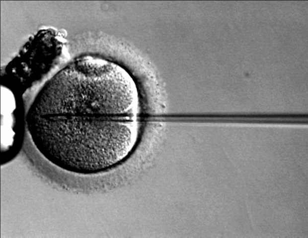
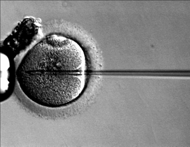
A needle being injected into a micro organism

Researchers at Boston Children’s Hospital report creating the first human tissue model of an inherited heart arrhythmia, replicating two patients’ abnormal heart rhythms in a dish, and then suppressing the arrhythmia with gene therapy in a mouse model. Their work, published in two papers in the July 30 print issue of the journal Circulation, opens the possibility of developing single-dose gene therapy treatments for inherited arrhythmias, and perhaps more common arrhythmias such as atrial fibrillation.


Our hope is to give gene therapy in a single dose that would work indefinitely. Our work provides proof-of-concept for a translatable gene therapy strategy to treat an inherited cardiac arrhythmia.”


Vassilios Bezzerides, MD, PhD, attending cardiologist in the Inherited Cardiac Arrhythmias Program at Boston Children’s Hospital


The two studies focused on catecholaminergic polymorphic ventricular tachycardia (CPVT), a leading cause of sudden death in children and young adults. The arrhythmia is typically triggered by exercise or emotional stress, and first becomes apparent at an average age of 12, often as a sudden loss of consciousness.


Current treatment consists of drugs such as beta-blockers and flecainide, surgery to disconnect nerves innervating the left side of the heart, an implanted cardioverter-defibrillator (which can lead to life-threatening complications in CPVT), and simply having children exercise as little as possible.


“Treatments for CPVT are currently pretty inadequate: 25 to 30 percent of patients will have recurrent life-threatening arrhythmias despite treatment,” says Bezzerides.


Building arrhythmic tissue


One study, published online by Circulation July 17, used tissue engineered models to investigate how CPVT works at the cellular and molecular level. It was led by William T. Pu, MD, of Boston Children’s Hospital and Kevin Kit Parker, PhD, of Boston Children’s and Harvard’s School of Engineering, Arts, and Sciences (SEAS).


Working with the Inherited Cardiac Arrhythmias Program, led by Dominic Abrams, MD, MBA, the researchers obtained blood samples from two patients at Boston Children’s Hospital who had CPVT caused by separate mutations in RYR2, the gene linked to most cases of CPVT. RYR2 encodes a channel that enables cells to release calcium — the first step in initiating a heart contraction.


The scientists then reprogrammed the patients’ blood cells to become induced pluripotent stem (iPS) cells, capable of making virtually all cell types. From these, they made cardiomyocytes (heart muscle cells) carrying the CPVT mutations and used them to construct models of heart-muscle tissue.


“The cells were seeded on an engineered surface so that they lined up in a specific direction similar to how heart muscle is organized,” explains Pu, who is director of Basic and Translational Cardiovascular Research at Boston Children’s. “The cells have very abnormal beating individually, but after assembly into tissue, they beat together, better modeling the actual disease. That’s why tissue-level models are important.”


Exercise test in a dish


Using a so-called optogenetic system, the team then applied blue light to one end of the tissue to activate the cells. This created an impulse that moved along the sheet of cells to produce a contraction. Using this system, they created an “exercise test in a dish.” To simulate exercise, they added the drug isoproterenol (similar to the stress hormone adrenaline) and applied infrared light to initiate faster heartbeats.


This testing helped reveal CPVT’s underlying mechanisms. When healthy heart tissue underwent the exercise test, calcium moved through the tissue in even waves. But in tissue models made from patients with CPVT, calcium waves moved at varying speeds, and in some parts of the tissue not at all, resulting in an abnormal circular motion known as re-entry — much like what happens in real life.


“When we paced the cells faster, the CPVT tissue sustained re-entrant arrhythmias, whereas normal tissue could handle it fine,” says Pu.


To understand how stress makes CPVT patients vulnerable to life-threatening arrhythmias, Pu, Parker, and colleagues identified signaling molecules that are activated by adrenaline, and then used drugs and CRISPR/Cas9 genome editing to selectively inhibit or modify them.


Through this strategy, they found that in healthy heart tissue, an enzyme called CaM kinase (CaMKII) chemically modifies RYR2, triggering the heart-muscle cells to release more calcium. In CPVT cells, this modification combines with the inherited RYR2 mutation to cause excessive calcium levels in cells, which precipitates arrhythmias.


“Nature designed CaMKII as part of the fight-or-flight response,” explains Pu. “When you get excited, you release more calcium so the heart can beat faster. But when RYR2 is mutated, the channel is leaky, so the cell releases way too much calcium, which causes arrhythmia.”


When the researchers blocked the CaMKII modification, they eliminated the arrhythmias in the tissue model. They got the same effect when they blocked CaMKII itself with the peptide AIP, a potent and selective CaMKII inhibitor.


“The coupling of iPS technology and organs on chips offers new opportunities for studies in precision medicine and the benefit of patients,” notes Parker. “Our vision is to use these technologies to screen patients with rare diseases for clinical trial enrollment. By replicating the patient’s disease in vitro, we can test candidate therapies on the patient’s disease and measure safety and efficacy, so that the right patients get tested with the right drug.”


Inhibiting CaMKII with gene therapy


Because the CaMKII enzyme acts on many tissues beside the heart — and is required by the brain for memory formation — the team wanted to be able to inhibit CaMKII in the heart specifically. In a separate study, published online by Circulation on June 3, a team led by Bezzerides and Pu tested a gene therapy approach in a mouse model of CPVT.


They engineered a special virus which, injected into mice with CPVT, selectively travelled to the heart and delivered AIP. Testing showed that AIP was expressed in about 50 percent of heart cells, enough to suppress arrhythmias, but not significantly expressed in non-heart tissues including the brain.


The researchers now plan to refine their gene therapy strategy and test it in a large animal model and eventually in patients with CPVT, likely in collaboration with other medical centers.


A general approach to heart disease?


Bezzerides and Pu believe the therapy could be effective for patients with CPVT caused by a variety of RYR2 mutations (more than 160 mutations have been reported). And they believe their overall strategy of inhibiting CaMKII in the heart could help treat more common causes of heart disease.


“CaMKII is not needed for normal heart function, but it becomes activated in many forms of heart disease,” says Pu. “In mouse models of many forms of heart disease, such as ischemic cardiomyopathy, atrial fibrillation, or hypertrophic cardiomyopathy, chronic CaMKII activation is detrimental. It is possible that our gene therapy approach to CaMKII inhibition could improve outcomes in these other types of heart disease.”

Being overweight around age 18 linked with higher risk of cardiomyopathy in adulthood

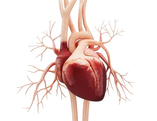
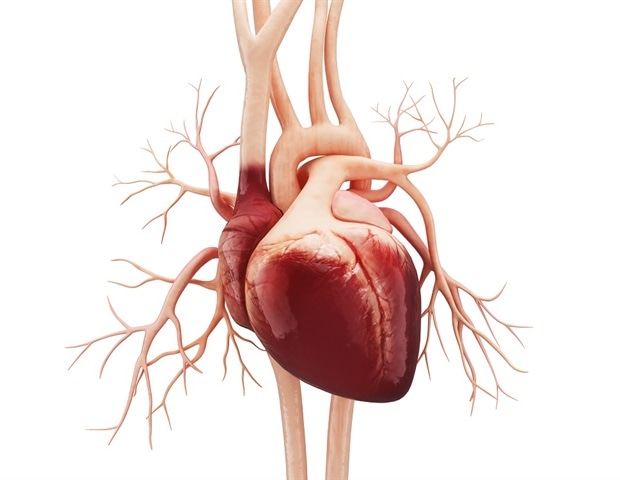
A human heart with all its veins with a white background

A large study of Swedish men found that those who were even mildly overweight around age 18 were more likely develop cardiomyopathy in adulthood — an uncommon heart muscle condition that can cause heart failure, according to new research in the American Heart Association’s journal Circulation.


The study examined data on height, weight and overall fitness from a Swedish registry of 1,668,893 men who enlisted in compulsory military service between 1969 and 2005, when the men were 18 or 19. The researchers then used two other national databases that track the causes of all hospitalizations and deaths in Sweden to determine whether the men had serious heart disease as they aged, and followed them for up to 46 years.


We were interested in studying cardiomyopathies, because heart failure caused by this historically uncommon disorder doubled in Sweden between 1987 and 2006.”


Annika Rosengren, M.D., Ph.D. study co-author, professor of medicine at the University of Gothenburg and cardiologist at Sahlgrenska University Hospital in Gothenburg, Sweden


Among the men in the study, 4,477 were diagnosed with cardiomyopathy at an average age of 45.5 years. The men who were lean at age 18 – with a body mass index (BMI – measure of body weight) below 20 – had a low risk of cardiomyopathy. However, that risk steadily increased as weight increased, even among men on the high end of a normal BMI (22.5-25).


There are several types of cardiomyopathy, but the causes are poorly understood. In one form, called dilated cardiomyopathy, the heart muscle becomes weak and can’t pump blood efficiently. In another, called hypertrophic cardiomyopathy, the heart muscle becomes stiff and isn’t able to fill with blood properly. Cardiomyopathy can reduce heart function and lead to heart failure, which means the heart is not able to pump blood properly.


In the study, men who had a BMI of 35 and over in their youth were eight times more likely to develop dilated cardiomyopathy as adults compared to men who were lean in their youth. It was not possible to estimate increased risk for hypertrophic cardiomyopathy in men with BMI 35 and above because there were too few cases to provide a meaningful analysis.


Because overall rates of cardiomyopathies are so low, it took a dataset as large and long-term as this one to have enough statistical power to find an association with weight, said Rosengren. She is not aware of other datasets large enough for a comparison study, but she would expect the results of this study to apply to men throughout the world, including the United States, although additional studies would need to be conducted to see if there are racial or ethnic differences in how body weight in adolescence influences the development of cardiomyopathy later in life.


The findings may or may not translate to women. The data on weight was gathered when males in Sweden register for compulsory military service. Since women do not register for military service, data on women’s weight at around age 18 was not available to the researchers.


New statement emphasizes need for special multidisciplinary clinical programs

Need for special multidisciplinary clinical programs

With a better understanding of how various heart conditions are inherited, and the availability of faster and less expensive genetic testing, there is need for more specialized multidisciplinary clinical programs that combine focused expertise in heart disease and genetics, according to a new statement from the American Heart Association, the world’s leading voluntary organization focused on heart and brain health. The statement is published in the Association’s journal Circulation: Genomic and Precision Medicine.


Cardiovascular genetics, as a subspecialty, has grown exponentially with the advances in genome sequencing and genetic testing and the growing understanding of the genetic basis of multiple heart conditions. Challenges exist with rapid growth, including the interpretation of genetic test results and the evaluation, counseling and management of genetically at-risk family members who have inherited the genetic alteration that increases their predisposition to a certain disease even if they have not yet shown signs or symptoms.


The state of genetic understanding of some heart disorders has improved to the point that we can use this information to help families and offer hope in ways never before possible. But it’s important that we have the right people, including medical geneticists and genetic counselors, as well as adequate facilities, equipment and other resources in place to provide clear and accurate guidance to these families throughout testing and decision-making processes.”


Ferhaan Ahmad, M.D., Ph.D., Chair of the Writing Group for the Statement, Associate Professor of Cardiovascular Medicine and Molecular Physiology and Director of the Cardiovascular Genetics Program with University of Iowa Health Care in Iowa City


The statement lays out what would be needed to create a quality specialized program:


  • Leadership from a cardiologist well-versed in genetics or a geneticist well-versed in cardiovascular medicine;
  • Core personnel including cardiologists, medical geneticists, genetic counselors, nurse managers and clinic coordinators;
  • Facilities for several types of cardiac imaging and people with the expertise to interpret the findings and recognize uncommon heart conditions;
  • The subspecialists and facilities to offer (either at the program or with a well-developed referral plan for complex surgeries) the invasive procedures necessary to diagnose and treat electrical problems in the heart, to assess heart function, to surgically correct structural problems of the heart, valves and aorta and to perform heart transplants;
  • Genetic testing and counseling; and
  • Support from other specialties such as sleep medicine, behavioral medicine, nutrition, social work and exercise physiology.


Currently, there are a few broad-based cardiovascular genetics programs in existence at academic centers, as well as smaller programs focused on a specific disease, such as hypertrophic cardiomyopathy — the most common genetic disorder of the heart, which results in a thickened heart muscle that has a harder time pumping blood.


Statement authors said a specialized genetic program that provides the integration of clinical cardiovascular findings – including those obtained from physical examination, imaging and functional assessment – with genetic information allows for improved diagnosis, prognosis and generational family testing to identify and manage risk and in certain cases to provide genotype-specific therapy.


Specialized programs in cardiovascular genetics could benefit both those with inherited heart conditions and their healthy family members, according to the statement.


“A patient who has a rare, genetic heart disease will benefit from specialized care from experts who know how to manage a disease that’s not familiar to the general cardiologist,” said Kiran Musunuru, M.D., Ph.D., M.P.H, associate professor of cardiovascular medicine and genetics at the University of Pennsylvania and outgoing editor-in-chief of Circulation: Genomic and Precision Medicine, who wasn’t involved in the development of the guideline. “Others having a close relative with an inherited heart disease can be screened to see if they have inherited genes that put them at risk for getting the disease in the future. If so, they can be monitored over time for early signs of disease and they can, in some cases, be treated to prevent the most serious consequences of the disease.”


Ahmad said in addition to providing quality care to families, these centers will also become key places for the genetics training of internal medicine and pediatric residents and cardiology fellows.


The American Heart Association is prepared to be part of the educational efforts needed to translate advances in genetics into better care for families with heart disease.


“The American Heart Association, led by the Council on Genomic and Precision Medicine and the Institute for Precision Cardiovascular Medicine, is primed and ready to support the education of clinicians and researchers in genetics and data science through the Precision Medicine Platform,” said Jennifer L. Hall, Ph.D., Chief of the American Heart Association’s Institute for Precision Cardiovascular Medicine. “We’re connecting clinicians and researchers by funding innovative data science, opening up new data sources with rich genetic information and allowing collaborators to analyze data in real time together on the Precision Medicine Platform in secure workspaces equipped with high performance computing and analytical tools.”


Other types of genetic diseases that might be diagnosed and treated at a specialized program include those that result in abnormally high levels of bad cholesterol, vascular disorders such as Marfan syndrome that can weaken the aorta, and abnormalities of heart rhythm that can raise the risk of sudden death. While cardiovascular genetics programs may serve both adults and children, these programs and the new scientific statement are unrelated to the treatment of congenital heart defects – structural birth defects of the heart that are not inherited.


Source:


American Heart Association


Journal reference:


Ahmad, F. et al. (2019) Establishment of Specialized Clinical Cardiovascular Genetics Programs: Recognizing the Need and Meeting Standards: A Scientific Statement From the American Heart Association. Circulation: Genomic and Precision Medicine. https://www.ahajournals.org/doi/abs/10.1161/HCG.0000000000000054.


Study finds promising new treatment for infants with Noonan Syndrome

An artificial heart kept on a doctors report

Noonan Syndrome (NS) is a rare genetic syndrome typically evident at birth and often linked to early-onset severe heart disease. NS is part of a group of diseases termed RASopathies that are caused by activating mutations of proteins belonging to the Ras and mitogen-activated protein kinase families.


In a new study, researchers at Université de Montréal and CHU Sainte-Justine Research Center show that a MEK inhibitor called trametinib can reverse hypertrophic cardiomyopathy (HCM) and valvular obstruction in patients with RIT1-associated NS. The groundbreaking research is published in the Journal of the American College of Cardiology.


”Up to this finding, our therapeutic options were limited to surgery, including heart transplant, and symptomatic relief with medication,” said the study’s author, Dr. Gregor Andelfinger, a pediatric cardiologist at CHU Sainte-Justine, a researcher at Sainte-Justine University Hospital Research Center in the fetomaternal and neonatal pathologies axis, and an associate research professor in the pediatrics department of Université de Montréal.


“Trametinib treatment is the first approach specifically targeted to the molecular cause of RASopathies,” said Dr. Andelfinger. “While our numbers are still very limited, we report the first patients in whom we were not only able to stabilize, but to reverse the disease of the heart. These results pave the way for larger trials, which are now needed.”


Dramatic improvement


Infants less than six months old with NS, HCM and congestive heart failure normally have a poor prognosis, with a one-year survival rate of 34 per cent. In the new study, the Sainte Justine clinical teams used trametinib, an inhibitor targeted specifically against the activating nature of the mutations, to try to treat NS in two patients.


They observed dramatic improvement of clinical and cardiac status in the patients only three months after treatment. Hypertrophy regressed in both patients, with sustained improvement over a total of 17 months of treatment, and normalization of laboratory values. One of the patients, who required ventilation, could be extubated after six weeks of treatment. Both patients showed better overall growth after treatment was started.


“The findings described in this report suggest that a life-threatening form of heart disease affecting young infants might be treatable, which, if true, would be unprecedented and so meaningful for the families whose lives this devastating problem touches,” commented Dr. Bruce Gelb, director of the Mindich Child Health and Development Institute at the Icahn School of Medicine at Mount Sinai, in New York City.


“Now we need to perform a proper clinical trial to prove that this drug is definitely working for this particular problem,” he said.


A promising first


Although the study was limited to two patients, given the promising results these outcomes suggest that MEK inhibition merits further study as a mechanistic treatment option for patients with RASopathies, the researchers believe. The study raises important questions for the treatment of such cases, in particular with respect to long-term efficacy and impact on other RASopathy manifestations.


Because of the role MEK plays in signaling heart growth, Gregor Andelfinger believes studies with a larger number of participants are now required to evaluate long-term side effects, optimal dosing and optimal treatment windows as well as investigate this treatment for other types of heart disease. It is conceivable that MEK inhibition may prove most effective during a fixed time window before the onset of irreversible cardiac remodeling in RASopathies, including those caused by genes other than RIT1.


Scientists aim to unlock the secrets of heart cells

A human heart with all its veins with a white background

The human heart appears to be pure physics: valves open and close, blood flows through the heart’s chambers, electrical impulses regulate the heartbeat. But a German-British research team is looking at it through a different perspective. It wants to break the heart down into its smallest parts – the cells – and unlock the secrets hidden there. The project is funded with €2.5 million over three years.


The scientists have the goal of understanding what causes the heart to become weak or sick. The project, which is coordinated at the Max Delbrück Center for Molecular Medicine in the Helmholtz Association (MDC, therefore focuses mainly on two forms of heart failure: dilative cardiomyopathy (DCM) and hypertrophic cardiomyopathy (HCM). In both conditions the heart is unable to pump blood properly, but for different reasons. In patients suffering from DCM the left ventricle is stretched out of shape like a wet sock, while HCM is characterized by a thickening of the walls of the ventricle. The researchers know that both processes cause changes to occur in the heart muscle cells and that connective tissue cells also play a role.


“We don’t yet have a good understanding of what exactly happens at the molecular level,” says Prof. Norbert Hübner, the project’s leader and a scientist at the Max Delbrück Center for Molecular Medicine in the Helmholtz Association (MDC). The research team therefore plans to use cutting-edge techniques to analyze thousands of individual cells from different parts of the heart, for example, to find out what genes and proteins are present.


No two heart cells are the same


This approach, which involves directly comparing cells from healthy and failing hearts, may lead to groundbreaking findings. “Our research could pave the way for creating novel therapies for heart failure or methods that prevent heart failure from developing in the first place,” says Hübner. Currently, there is no causal treatment and thus no cure. Each year some 200,000 patients with heart failure are admitted to hospitals, and those with the most severe cases wind up on the transplant list.


Even though single-cell analysis holds the promise of exciting discoveries, researchers can only give a correct description of a cell population because cells are influenced by a wide range of factors in their environment. These include not only pressure in the ventricles, but also interactions with other cell types, neurotransmitters, and immune processes. “No two cells are alike, not even healthy heart cells,” explains Hübner. The spatial localization of cells and the computer modeling of cell populations are therefore incorporated into the research strategy.


The samples are provided by the British partners


The German and British researchers complement each other in the valuable resources and expertise that they bring to the project. The British project partners have at their disposal human heart samples obtained from healthy and diseased donors. Thomas Eschenhagen’s team in Hamburg knows how to cultivate heart tissue in the lab. This will enable the researchers to model heart diseases and test pharmacological agents, which could lead to the discovery and development of new drugs.


The project – entitled “Spatially resolved cellular and molecular drivers of cardiac remodeling in healthy and failing” – is receiving €2.5 million in total funding over three years from the German Center for Cardiovascular Research (DZHK) and the British Heart Foundation (BHF).