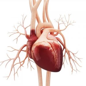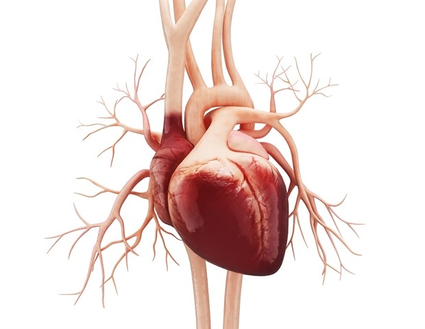
The human heart appears to be pure physics: valves open and close, blood flows through the heart’s chambers, electrical impulses regulate the heartbeat. But a German-British research team is looking at it through a different perspective. It wants to break the heart down into its smallest parts – the cells – and unlock the secrets hidden there. The project is funded with €2.5 million over three years.
The scientists have the goal of understanding what causes the heart to become weak or sick. The project, which is coordinated at the Max Delbrück Center for Molecular Medicine in the Helmholtz Association (MDC, therefore focuses mainly on two forms of heart failure: dilative cardiomyopathy (DCM) and hypertrophic cardiomyopathy (HCM). In both conditions the heart is unable to pump blood properly, but for different reasons. In patients suffering from DCM the left ventricle is stretched out of shape like a wet sock, while HCM is characterized by a thickening of the walls of the ventricle. The researchers know that both processes cause changes to occur in the heart muscle cells and that connective tissue cells also play a role.
“We don’t yet have a good understanding of what exactly happens at the molecular level,” says Prof. Norbert Hübner, the project’s leader and a scientist at the Max Delbrück Center for Molecular Medicine in the Helmholtz Association (MDC). The research team therefore plans to use cutting-edge techniques to analyze thousands of individual cells from different parts of the heart, for example, to find out what genes and proteins are present.
No two heart cells are the same
This approach, which involves directly comparing cells from healthy and failing hearts, may lead to groundbreaking findings. “Our research could pave the way for creating novel therapies for heart failure or methods that prevent heart failure from developing in the first place,” says Hübner. Currently, there is no causal treatment and thus no cure. Each year some 200,000 patients with heart failure are admitted to hospitals, and those with the most severe cases wind up on the transplant list.
Even though single-cell analysis holds the promise of exciting discoveries, researchers can only give a correct description of a cell population because cells are influenced by a wide range of factors in their environment. These include not only pressure in the ventricles, but also interactions with other cell types, neurotransmitters, and immune processes. “No two cells are alike, not even healthy heart cells,” explains Hübner. The spatial localization of cells and the computer modeling of cell populations are therefore incorporated into the research strategy.
The samples are provided by the British partners
The German and British researchers complement each other in the valuable resources and expertise that they bring to the project. The British project partners have at their disposal human heart samples obtained from healthy and diseased donors. Thomas Eschenhagen’s team in Hamburg knows how to cultivate heart tissue in the lab. This will enable the researchers to model heart diseases and test pharmacological agents, which could lead to the discovery and development of new drugs.
The project – entitled “Spatially resolved cellular and molecular drivers of cardiac remodeling in healthy and failing” – is receiving €2.5 million in total funding over three years from the German Center for Cardiovascular Research (DZHK) and the British Heart Foundation (BHF).