WSU research sheds new light on a drug that may be used to treat hypertrophic cardiomyopathy

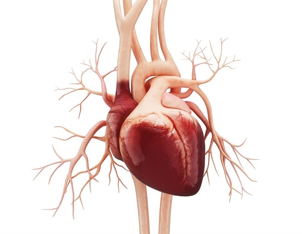
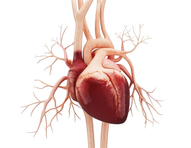
A red artificial heart kept on a medical paper

Research at Washington State University sheds new light on one molecule that may be used to treat a heart condition that can lead to stroke, heart attack and other forms of heart disease.

That molecule is mavacamten. Scientists at WSU’s Integrative Physiology and Neuroscience department discovered it suppresses excessive force generated by hyper-contractile muscle cells in the human heart.

The research, published in the British Journal of Pharmacology, is especially significant for those with hypertrophic cardiomyopathy, a genetic condition where the left ventricle wall of the heart is enlarged. If left untreated, hypertrophic cardiomyopathy can lead to cardiac fibrosis, stroke, heart attack, heart failure, other forms of heart disease and a condition known as sudden arrhythmic death syndrome.

Too much contraction leads to thicker, stiffer hearts, where the heart contracts so much it is unable to properly fill with blood as the heart relaxes. This ends up pushing less blood out of the heart with each heartbeat and, in turn, less blood pumped throughout the body like it is supposed to be.”

Peter Awinda, first author on the paper and scientific manager in Bertrand Tanner’s laboratory at WSU

Hypertrophic cardiomyopathy affects men and women equally. About 1 out of every 500 people have the disease.While there are some genetic markers to detect it, most people only discover their condition after a cardiac event that often results in a hospital visit.

The research

The project is a collaboration between the Tanner Laboratory in Pullman and Ken Campbell’s laboratory at University of Kentucky. Campbell manages a human cardiac biobank, where he ships tissue samples frozen in liquid nitrogen to Tanner, who is the principal investigator for the research.

After arriving in Pullman, the cardiac tissue was thawed, ‘skinned’ to remove the cell membrane, and trimmed to the right dimensions for an experiment.

Three micrograms of the drug, mavacamten, were then applied to some of the prepared tissue samples; other samples did not receive the drug and were labeled as controls.

To activate muscle contraction Awinda applied calcium to the tissue.

“As we increase calcium concentration it encourages contraction and the muscle goes from relaxed to contracted, and so we were testing the drug against these different levels of force,” Awinda said.

He found the drug reduces the maximal force of contraction by nearly 20 to 30% compared to the controls.

“The drug is successful because it is an inhibitor of myosin, which is one of the proteins required for the muscle contraction process,” Awinda said. “The research shows this could be a good candidate to treat hypertrophic cardiomyopathy.”

The collaborative study was made possible by organ donors and their families. The work was paid for by a $300,000 grant from the American Heart Association to Tanner and Campbell.

These initial studies helped Tanner and Campbell add an additional $2.8 million grant from the National Institutes of Health to support additional work in this area over the next 4 years.

Next steps

One of the research team’s next goals is to see how mice with a human mutation for hypertrophic cardiomyopathy respond to the drug.

Awinda said researching mice expressing the human gene is significant because it may provide a connection to what is seen in humans.

“When we see the same effects in the mice samples that we see in the human tissues, the drug is doing what it is intended to,” he said. “Both studies really help reinforce our understanding and inform us of what we see happening.”

Journal reference:

Awinda, P.O., et al. (2020) Effects of mavacamten on Ca2+ sensitivity of contraction as sarcomere length varied in human myocardium. British Journal of Pharmacology. doi.org/10.1111/bph.15271.

Cats’ immune system can deal with SARS-CoV-2, shows study

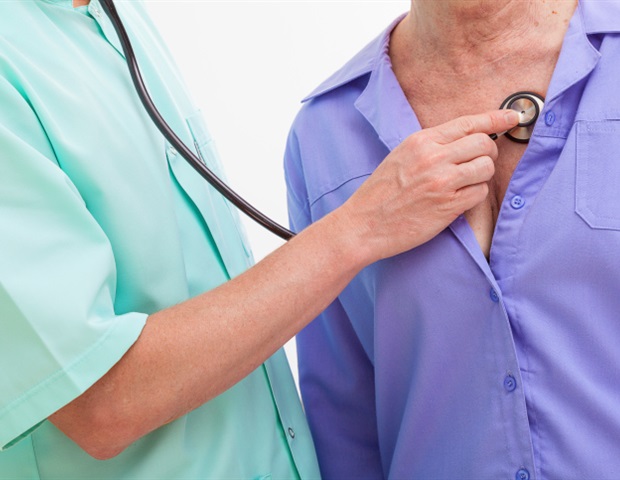
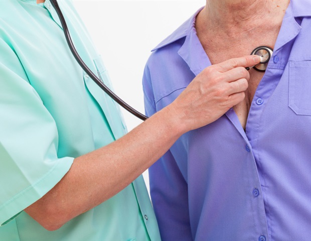
A doctor checking a patient with his stethoscope

On 8 May 2020, the Institute of Agrifood Research and Technology (IRTA) reported the case of the first cat infected with SARS-CoV-2 in Spain. It was a 4-year-old cat called Negrito, who lived with a family affected by COVID-19, with one case of death.

Coinciding with these facts, the animal presented severe respiratory difficulties and was taken to a veterinary hospital in Badalona (Barcelona), where it was diagnosed with hypertrophic cardiomyopathy. Due to a terminal condition the hospital decided to do a humanitarian euthanasia.

The necropsy, performed at the High Biosafety Level Laboratories of the Animal Health Research Center (CReSA) at IRTA, confirmed that Negrito suffered from feline hypertrophic cardiomyopathy and had no other lesions or symptoms compatible with a coronavirus infection.

The RT-PCR test confirmed that the animal had become infected with SARS-CoV-2, but with a very low and residual viral load.

To date there have been few cases of feline SARS-CoV-2 infection worldwide, which is why researchers have deepened the case study and published it recently in the journal Proceedings of the National Academy of Sciences (PNAS).

They performed serological tests on the cat Negrito and another cat that also lived in the same home, Whisky, which did not show any signs of disease. The tests, carried out by the AIDS Research Institute (IrsiCaixa), show that the two cats had developed antibodies against SARS-CoV-2.

“In both cases we have detected neutralizing antibodies, in other words, they have the ability to bind to the virus and block it,” explains Julià Blanco, IGTP researcher at IrsiCaixa.

He goes on to say “this is important because it shows us that the immune system of cats can deal with SARS-CoV-2 and, in these specific cases, protect them from developing symptoms”.

Experimental studies currently being conducted show that cats, in addition to becoming infected with SARS-CoV-2, can transmit it to other nearby cats, but without any clinical signs.

However, the first suspicions of researchers were that both Negrito and Whisky had been infected by their owners because they had not had contact with other cats.

To check this, the team analyzed the genetic sequence of the virus that Negrito had and found that “it has a 99.9% similarity to the virus of the owner who died, this suggests that the cat became directly infected from family members”, explains Marc Noguera-Julián, researcher at IrsiCaixa.

Given the number of people infected worldwide and the few reported cases of animals, experts continue to note that “pets play a negligible role in the epidemiology of SARS-CoV-2 and, in particular, cats become very residually infected and there is no evidence of transmission of the virus to humans. This is a case of reverse zoonosis, in which cats are the side victims without the virus causing them health problems”, states Júlia Vergara-Alert, researcher at IRTA-CReSA.

So far, there has only been one reported episode in the Netherlands in which a farmer became infected through minks, which would be the first known potential case of COVID-19 zoonosis.

IRTA and the Council of Veterinary Associations of Catalonia, with the collaboration of the Veterinary Clinical Hospital of the Universitat Autònoma de Barcelona are studying whether pets can become infected with SARS-CoV-2

Following the Negrito case, in May the IRTA-CReSA coronavirus research team, in coordination with the Council of Colleges of Veterinarians of Catalonia and the Veterinary Clinical Hospital of the Universitat Autònoma de Barcelona, started a study to assess how often cats, dogs and ferrets have become infected with SARS-CoV-2 from people in a family setting.

The study is being carried out with the voluntary participation of veterinary clinics and hospitals in Catalonia and consists of taking samples of oropharyngeal and rectal swabs, as well as animal serum.

We want to check if the case of Negrito and Whisky has been punctual or is repeated in more cases; in this way we will have more scientific information on how the most common pets can become infected with SARS-CoV-2 and to what degree”,

Joaquim Segalés, IRTA-CReSA Researcher and Professor, Faculty of Veterinary Medicine, The Autonomous University of Barcelona

The professor also remarks that “so far, it is known that around the world there have been some cases of cats and dogs that have become infected through their owners, but these animals have not suffered a serious illness and there is no evidence that they have transmitted the virus to their owners”.

Journal reference:

Segalés, J., et al. (2020) Detection of SARS-CoV-2 in a cat owned by a COVID-19−affected patient in Spain. Proceedings of the National Academy of Sciences. doi.org/10.1073/pnas.2010817117.

Study: Mavacamten improves heart function in patients with hypertrophic cardiomyopathy

A woman instruction other women in a swimming pool

Mavacamten improves heart function and symptoms in patients with obstructive hypertrophic cardiomyopathy, according to results of the EXPLORER-HCM trial presented in a Hot Line session today at ESC Congress 2020.

The results of this pivotal trial support a role for disease-specific therapy for obstructive hypertrophic cardiomyopathy (HCM) which treats the cause instead of just managing symptoms.

Iacopo Olivotto, Professor and Principal Investigator, Careggi University Hospital

HCM affects approximately 1 in 500 people. It is defined by left ventricular hypertrophy that cannot be explained by another cardiac or systemic disease.

The majority of HCM patients have obstructive HCM, where a combination of cardiac hypertrophy, excess contractility and abnormal movement of the mitral valve blocks or reduces blood flow from the left ventricle to the aorta – called left ventricular outflow tract (LVOT) obstruction.

Common symptoms include dyspnoea, atypical chest pain, palpitations, fatigue, and feeling lightheaded or fainting. Some people have few or no symptoms.

But for others, HCM is a debilitating and life-changing disease resulting in physical limitations and lower quality of life. In some patients, the left ventricular remodelling progresses to refractory heart failure.

Currently available medical treatments focus on symptom relief and fail to address the underlying causes of obstructive HCM. These non-specific agents often have modest efficacy or substantial side effects.

Surgical septal myectomy and alcohol septal ablation are efficacious but carry the risks inherent to invasive procedures and require specific expertise that is not always available. Therefore, an effective pharmacological therapy for obstructive HCM is an important unmet need.

Mavacamten is a first-in-class cardiac myosin inhibitor that directly targets the underlying pathophysiology of HCM and restores the heart’s normal function.

In early clinical trials, treatment with mavacamten led to significant improvements of symptoms, physical function, exercise capacity, and quality of life, and reduced LVOT obstruction in patients with obstructive HCM.

EXPLORER-HCM was a pivotal, global, phase 3, randomised, placebo-controlled clinical trial that tested the efficacy and safety of mavacamten in treating symptomatic obstructive HCM. A total of 251 patients received once daily mavacamten or placebo for 30 weeks. The endpoints were chosen to examine exercise capacity, symptoms, LVOT obstruction, functional status, and quality of life.

The primary endpoint assessed the treatment effect of mavacamten at week 30 relative to placebo on both symptoms and cardiac function.It was defined as achieving 1) ≥1.5 mL/kg/min improvement in peak oxygen consumption (peak VO2) and ≥1 New York Heart Association (NYHA) class reduction OR 2) ≥3.0 mL/kg/min improvement in peak VO2 and no worsening of NYHA class.

Secondary endpoints included change from baseline to week 30 in post-exercise LVOT gradient and patient-reported outcomes such as the Kansas City Cardiomyopathy Questionnaire-Clinical Summary Score (KCCQ-CSS) and HCM Symptom Questionnaire-Shortness-of-Breath (HCMSQ-SoB) subscore.

At week 30, 45 (36.6%) patients on mavacamten met the primary composite endpoint versus 22 (17.2%) on placebo (p=0.0005). All secondary endpoints, including post-exercise LVOT gradient and patient-reported outcomes, also demonstrated statistically significant improvements for mavacamten as compared to placebo (all p<0.0006).

Safety and tolerability with mavacamten were similar to placebo. Some 11 serious adverse events were reported in 8.1% of patients on mavacamten versus 20 events in 8.6% of patients on placebo.

Serious cardiac adverse events occurred in four patients treated with mavacamten (two atrial fibrillation, two stress cardiomyopathy), and four in the placebo group (three atrial fibrillation, one atrial fibrillation and congestive heart failure).

Professor Olivotto said: “The totality and consistency of the results showed benefit of mavacamten treatment compared to placebo in patients on background HCM therapy. Mavacamten improved functional capacity, LVOT gradient, symptoms, and key aspects of quality of life in patients with obstructive HCM and was generally well tolerated.”

New study assesses cardiac problems in elite female athletes

New study assesses cardiac problems in elite female athletes

The first-ever cardiac study of elite female basketball players in the United States shows how their hearts adapt to intense physical training.

The study of 140 WNBA players also provides physicians with a frame of reference when screening for cardiac problems in female athletes.

The research is the result of a long-standing collaboration among cardiologists and researchers at Columbia University Irving Medical Center and NewYork-Presbyterian, the National Basketball Association and Women’s National Basketball Association, and the National Basketball and Women’s National Basketball Players Associations that has also created a screening and monitoring program for the leagues and guidelines for NBA and WNBA team physicians. The research team has previously reported similar data on male NBA players.

The new research, published in JAMA Cardiology, also suggests that similar studies of female athletes are needed for other sports, which affect the heart in different ways.

Why study WNBA players?

Intense physical exercise increases the size, mass, and thickness of the walls of the heart.

“We call this ‘athlete’s heart,’ and we’ve recognized this remodeling process for decades,” says David J. Engel, MD, a sports cardiologist at NewYork-Presbyterian/Columbia University Irving Medical Center and senior author of the paper.

But nearly all published data on this describes athletic changes in the hearts of male athletes, and there is very little data on what an athletic heart looks like in elite female athletes and absolutely no data on the athletic heart in women who are professional basketball players.”

David J. Engel, MD, Study Senior Author and Sports Cardiologist, NewYork-Presbyterian/Columbia University Irving Medical Center

Team physicians and physicians who care for athletes need to know what a healthy heart in elite athletes looks like so that healthy hearts can be distinguished from those with signs of cardiac problems, including hypertrophic cardiomyopathy, an enlargement, and thickening of the heart that can lead to life-threatening arrhythmias.

Among U.S. athletes, basketball players have been shown in epidemiologic studies to have the highest incidence of exercise-induced sudden cardiac death, though the incidence is lower for female athletes than for males.

In 2006, the NBA was the first professional sports league to implement a standardized preseason screening program that tests all of its players for heart disease.

“But without normative data on how training for professional basketball affects heart structure compared with non-athletes or athletes from other sports, it isn’t easy to identify who is at risk,” says Sofia Shames, MD, a cardiologist at NewYork-Presbyterian/Columbia University Irving Medical Center and the study’s lead author.

Basketball strengthens heart

In the study, the researchers analyzed cardiac ultrasound data from the 2017 season in 140 WNBA athletes, all of whom had similar training.

The researchers found that the WNBA players generally have larger and thicker (more muscular) hearts than most women who are not athletes, and the differences can be attributed to a combination of the larger height of WNBA players and their physical conditioning.

According to heart disease screening guidelines, which are based on data from the general population, approximately 16% of the WNBA players could be inappropriately classified as having left ventricular enlargement.

“The general population is not particularly athletic, and a physician who sees a heart the size of many WNBA players’ hearts would do further testing,” Shames says.

“But in this group of women, these larger hearts are within the normal range for the athletes’ size and conditioning.”

“Evaluating the WNBA players on a large scale allows us to have comparison data for the future. Up until this research, some players were being sidelined until further tests could be done,” says Marci Goolsby, MD, team physician for the WNBA New York Liberty team, sports medicine physician at the Women’s Sports Medicine Center of the Hospital for Special Surgery, and a co-author of the paper.

“We now have a much clearer picture of how to interpret data from preseason echocardiograms, and we can better determine who requires further testing.”

The hearts of the WNBA players had also strengthened in different ways than the hearts of other female athletes. More than half of the WNBA players had heart walls greater than 1 cm in thickness, compared with less than 10% of Italian female athletes from a variety of sports.

The scans also showed that an important index of heart geometry — the thickness of the heart muscle in relation to diameter — was more prominent in the WNBA players compared to other female athletes.

“That’s why we need reference data for players in each specific sport,” Engel adds.

“If WNBA team physicians relied on the data from the European studies to screen players for heart disease, they would still be left with clinical questions. Our research can hopefully serve as a stimulus for more studies of female athletes in other sports.”

Journal reference:

Shames, S., et al. (2020) Echocardiographic Characterization of Female Professional Basketball Players in the US. JAMA Cardiology. doi.org/10.1001/jamacardio.2020.0988.

New mathematical model to study the behavior of the human heart

New model to study the behavior of the human heart

Having now reached its third year, Project “iHEART” of Politecnico di Milano, winner of an ERC Advanced Grant of euro 2,350,000, has set itself the ambitious goal of creating a complete mathematical model to study the behavior of the human heart and of its pathologies, a sort of “virtual microscope.”

This integrates all cardiac function processes, namely electrical impulse propagation, cellular activation, contraction and myocardial relaxation during the systolic and diastolic phase, blood fluid dynamics in both ventricles and atria, and opening-closure dynamics of the four cardiac valves.

Research carried out to date has already allowed to initiate operations of certain subproblems, which are attracting considerable interest in the medical community. Hence, from feasibility studies, focus has shifted to early field tests, in which mathematicians and doctors cooperate to optimise these new tools in the framework of delicate surgical procedures intended to solve certain very important cardiac conditions.

Some examples are given below.

Models developed by project iHeart have led to the production of quantitative indications on factors that contribute to trigger and maintain arrhythmias, such as ventricular tachycardia. Traditional surgical methods consist in performing transcatheter ablations, which allow, via radio frequencies, to deactivate the abnormal areas causing the arrhythmia.

In partnership with the Arrhythmology Unit and with the Cardiac Electrophysiology Unit of the IRCCS [scientific institute for hospitalisation and care] San Raffaele Hospital, Milan, researchers verified how cardiac mathematics can underpin and consolidate electrophysiological study in the localisation of intervention areas on the heart wall.

Increasingly rapid algorithms, which will allow to perform this type of analysis in real time, thus significantly speeding up the decisional process related to surgery, have also reached an advanced phase of development.

A model is currently being developed in partnership with the Sacco Hospital, Milan, to provide precise indications for the heart surgeon on how to perform myectomy (removal) of a portion of the interventricular septum using a very low cost, non-invasive analysis.

This treatment is the one that is most widely used to treat hypertrophic obstructive cardiomyopathy, which consists in a septal thickening which makes hard to eject blood from the left ventricle into the ascending aorta.

The mathematical simulation is inserted into the preoperative phase and was considered by medical doctors as an effective guiding tool for this surgical operation.

An additional computational tool has been developed in partnership with the Cardiology and Radiology Departments of S. Maria del Carmine Hospital, Rovereto (TN), to improve cardiac resynchronisation therapy (CRT), which consists in implanting a device capable of restoring correct synchronisation of the heart contraction impaired either by conduction disorders or by the presence of scars.

To this end, cardiologists have to map the left ventricle to detect its electrical activity by inserting a catheter-electrode into the blood vessels.

The currently validated mathematical instrument will allow to considerably reduce the mapping duration and, thus, the patient exposure to invasive treatment, besides guiding catheter placement in the most curative position for the patient presenting heart failure.

As shown by these examples, iHEART has opened new horizons between mathematics and translational medicine, and has established coordinated and systematic action between Universities and hospitals, creating a new professional figure at the interface between mathematics, bioengineering, medicine and data science.

As a result of all the new clinical partnerships, and the integrated activity of our young researchers (PhD students and post-doctorate graduates) with that of hospital-based researchers, we shall pave the way for a new discipline, namely Computational Medicine.”

Alfio Quarteroni, Professor and Project Manager, Politecnico di Milano

Alfio Quarteroni Professor, Politecnico di Milano, Milan, Italy and Professor Emeritus, EPFL, Lausanne, Switzerland acknowledged as one of the most multi-faceted mathematicians in the world, famous for applying mathematics to the most diverse fields: aerospace industry, environmental pollution, impact of earthquakes on civilian buildings, urban planning, medicine, and even competitive sports.

Particularly, he participated in producing the Solar Impulse aircraft, and directed the team of researchers who developed the mathematical model for Alinghi, the Swiss yacht twice winner of the prestigious America’s Cup, the Sailing Cup, for two consecutive editions of the race, in 2003 and 2007.

Reductive stress is intolerable, can induce heart failure

Reductive stress is intolerable, can induce heart failure

Many heart diseases are linked to oxidative stress, an overabundance of reactive oxygen species. The body reacts to reduce oxidative stress — where the redox teeter-totter has gone too far up — through production of endogenous antioxidants that reduce the reactive oxygen species. This balancing act is called redox homeostasis.

But what happens if the redox teeter-totter goes too far down, creating antioxidative stress, also known as reductive stress? Rajasekaran Namakkal-Soorappan, Ph.D., associate professor in the University of Alabama at Birmingham Department of Pathology, and colleagues have found that reductive stress, or RS/AS, is also pathological. This discovery, they say, may have clinical importance in management of heart failure.

They report that RS causes pathological heart enlargement and diastolic dysfunction in a mouse model. This study, published in the journal Antioxidants and Redox Signaling, was led by Namakkal-Soorappan and Pei Ping, Ph.D., David Geffen School of Medicine at the University of California-Los Angeles.

Antioxidant-based therapeutic approaches for human heart failure should consider a thorough evaluation of antioxidant levels before the treatment,” they said. “Our findings demonstrate that chronic RS is intolerable and adequate to induce heart failure.”

The study used transgenic mice that had upregulated genes for antioxidants in the heart, which increased the amounts of antioxidant proteins and reduced glutathione, creating RS. One mouse line had low upregulation, and one had high upregulation, creating chronic low RS and chronic high RS, respectively, in the hearts of the mice.

The mice with high RS showed pathological heart changes called hypertrophic cardiomyopathy, and had an abnormally high heart ejection fraction and diastolic dysfunction at 6 months of age. Sixty percent of the high-RS mice died by 18 months of age.

The mice with low RS had normal survival rates, but they developed the heart changes at about 15 months of age, suggesting that even moderate RS can lead to irreversible damage in the heart over time.

Giving high-RS mice a chemical that blocked biosynthesis of glutathione, beginning at about 6 weeks of age, prevented RS and rescued the mice from pathological heart changes.

Gobinath Shanmugam, Ph.D., postdoctoral fellow in the UAB Department of Pathology, and Namakkal-Soorappan point out that a 2019 survey found about 77 percent of Americans are consuming dietary supplements every day, and within this group, about 58 percent are consuming antioxidants as multivitamins. Thus, a chronic consumption of antioxidant drugs by any individual without knowing their redox state might result in RS, which can induce pathology and slowly damage the heart.

Effect of RS on skeletal muscle

In a related study, published in the journal Redox Biology, Namakkal-Soorappan looked at the impact of RS on myosatellite cells, which are also known as muscle stem cells. These cells, located near skeletal muscle fibers, are able to regenerate and differentiate into skeletal muscle after acute or chronic muscle injury. The regulation of myosatellite cells is of interest given the loss of skeletal muscle mass during aging or in chronic conditions like diabetes and AIDS.

Recently, Namakkal-Soorappan reported that tilting the redox teeter-totter to oxidative stress impaired regeneration of skeletal muscle. Now, in the Redox Biology paper, he has shown that tilting the redox to RS also causes significant inhibition of muscle satellite cell differentiation.

Rather than genetic manipulation to induce RS, as was done in the heart study, the researchers used the chemical sulforaphane or direct augmentation of intracellular glutathione to induce RS in cultured mouse myoblast cells. Both treatments inhibited myoblast differentiation. Finally, authors attempted to withdraw antioxidative stress by growing cells in medium without sulforaphane, which removes the RS and accelerates the differentiation. Namakkal-Soorappan and colleagues found that a pro-oxidative milieu, through a mild generation of reactive oxygen species, was required for myoblast differentiation.

The researchers also showed that genetic silencing of a negative regulator of the antioxidant genes also inhibited myoblast differentiation.

Journal reference:

Shanmugam, G., et al. (2020) Reductive Stress Causes Pathological Cardiac Remodeling and Diastolic Dysfunction. Antioxidants & Redox Signaling. doi.org/10.1089/ars.2019.7808.

ECG screening more effective at detecting cardiac conditions in college athletes

ECG detecting cardiac problems in college athletes

Over the past 30 years, colleges and universities have increasingly screened athletes for health conditions that may pose undue risk to sports participation. Sudden cardiac death is the leading cause of death among college athletes, so a primary function of these screenings is to reveal unknown heart conditions.

The National Collegiate Athletic Association requires a baseline pre-participation physical exam and patient history; some schools also administer an electrocardiogram (ECG), which measures the electrical output of a person’s heartbeat. Disagreement exists, however, about ECGs’ value. While experts generally agree that an ECG can flag cardiac issues of concern, detractors say they result in too many false-positive findings, leading to unnecessary additional tests and costs.

Research published today indicates that screenings that incorporate an ECG are more effective at detecting cardiac conditions that put athletes at risk, and more efficient in terms of cost-per-diagnosis of at-risk players, than screenings involving only a physical exam and patient history.

The journal Heart Rhythm published the multi-institutional research. It was led by Dr. Kimberly Harmon, section head of sports medicine at the University of Washington School of Medicine and lead physician for the UW football team.

Harmon and colleagues found that, across 8,602 records of Pacific-12 Conference athletes, cardiovascular screenings with an ECG in addition to a patient history and physical exam were six times more likely to detect a risky heart condition than screenings that involved only a patient history and physical. In parallel, ECG use improved the cost efficiency per diagnosis by five-fold.

This is a real-world assessment of the discoveries resulting from different screening strategies, and how much they cost. This study shows that screening with ECG is not only significantly more effective, it is only incrementally more expensive overall and costs much less per diagnosis. It can be implemented for much less than the cost of a pair of athletic shoes.”

Dr. Kimberly Harmon, section head of sports medicine at the University of Washington School of Medicine and lead physician for the UW football team

The researchers sought to compare positive findings, disease prevalence, and the costs of two screening strategies. They collected nine years of athletes’ de-identified data from Pac-12 Conference institutions, which employ varied strategies for pre-participation physical screenings. Also analyzed were secondary tests, cardiac diagnoses, return-to-play outcomes, and complications from testing.

From 8,602 screenings, 11 athletes were identified with cardiovascular conditions:

  • History and physical exam: Two athletes (of 4,955) were identified as at risk, equating with a prevalence of 0.05% (1 in 2,454). Per-athlete screening cost averaged $130; cost per diagnosis was $312,407.
  • History, physical and ECG: nine athletes (of 3,647) were identified as at risk, equating with a prevalence of 0.024% (1 in 410). Per-athlete screening test cost was $152; cost per diagnosis was $61,712.

The costs of screenings and secondary testing were based on the Centers for Medicare and Medicaid Services Physician Fee Schedule.

“This is the first study to compare screenings by using real-life outcomes rather than theoretical models and variable assumptions,” said Harmon, a UW professor of family medicine and of orthopedics and sports medicine.

Not every school can incorporate ECG tests into athlete screenings, Harmon said. It’s less a question about covering a $22 test cost and more about whether physicians are readily available to interpret the ECGs.

The study included screenings of athletes across all varsity sports, though men’s basketball and football account for more than 50% of sudden cardiac deaths among college athletes, she said. Several Pac-12 schools have prioritized those sports for screening.

The research was supported with a grant from the Pac-12 Conference’s Student-Athlete Health and Well-Being Initiative.

Journal reference:

Harmon, K.G, et al. (2020) Comparison of Cardiovascular Screening in College Athletes by History and Physical Examination with and without an Electrocardiogram: Efficacy and Cost. HeartRhythm. doi.org/10.1016/j.hrthm.2020.04.032.

Study shows why people with certain heart diseases may be more susceptible to COVID-19

certain heart diseases may be more susceptible to COVID 19

People with certain heart diseases may be more susceptible to worse outcomes with COVID-19, but the reason why has remained unknown.

New research from Mayo Clinic indicates that in patients with one specific type of heart disease obstructive hypertrophic cardiomyopathy (HCM) the heart increases production of the ACE2 RNA transcript and the translated ACE2 protein.

Normally, this pathological response at the cellular level might be the heart’s attempt to compensate for changes caused by disease. Unfortunately, SARS-CoV-2, the virus that causes COVID-19, hijacks these ACE2 receptors on the membrane of cells and uses them to get inside the cells.

The virus not only gains entry through ACE2, but also it takes this protein with it, removing a protective signaling pathway that normally counters the negative impact of the hormone angiotensin II. This hormone increases blood pressure and leads to fluid retention.

Over the course of a nearly 20-year study published in Mayo Clinic Proceedings, researchers analyzed frozen samples of heart muscle tissue from 106 patients who had surgery for obstructive hypertrophic cardiomyopathy. The control group used heart tissue from 39 healthy donor hearts.

Of all the RNA transcripts in the entire human genome, our research revealed that the single most upregulated RNA transcript in the heart muscle was ACE2. In fact, we confirmed a fivefold increase in ACE2 protein levels in the heart muscle of these patients with obstructive HCM. This could connect the dots and potentially explain why patients with certain heart diseases might fare worse with COVID-19.”

Michael Ackerman, M.D., Ph.D., Genetic Cardiologist, Mayo Clinic

Dr. Ackerman is director of the Windland Smith Rice Sudden Death Genomics Laboratory at Mayo Clinic and senior author on the study. This study involved national and international investigators.

The next step is to look for other elevated ACE2 levels by analyzing available heart tissue from patients who have died from hypertension and other heart diseases. Lung tissue from COVID-19 victims also could be analyzed to see if ACE2 levels are higher than in normal lung tissue.

“This discovery provides another reason for patients taking angiotensin-converting enzyme (ACE) inhibitors and angiotensin II receptor blockers to stay on their heart medications, as recommended by all major cardiac societies,” says Dr. Ackerman. “Removing these medications in a patient whose heart has elevated protein levels of ACE2 could cause even more tissue damage.”

Study identifies a protein and signaling pathway that may contribute to sudden cardiac death

heart disease written on a paper with red capsules kept on

By studying the sick hearts removed from four patients undergoing heart transplants, researchers have identified a protein and a signaling pathway that may contribute to sudden death in an inherited form of heart disease.

The patients were receiving new hearts to treat Arrhythmogenic Right Ventricular Cardiomyopathy (ARVC), a rare genetic disease that causes abnormal heart rhythms (arrhythmias) and increases the risk of sudden cardiac death, particularly during exercise or emotional excitement. ARVC affects between 1 in 1,000 and 1 in 1,250 people and is a leading cause of sudden death among young athletes who have no prior symptoms or cardiovascular disease diagnosis.

“We have identified a new pathway in heart cells that explains how arrhythmias occur in patients with ARVC,” says Long-Sheng Song, MD, professor of internal medicine at the University of Iowa Carver College of Medicine and one of the leaders of the study team that included researchers from the UI and the Fuwai Hospital, Chinese Academy of Medical Sciences and Peking Union Medical College in Beijing, China. “We hope that new drugs targeting this pathway can now be developed, which might help us treat this devastating, progressive disease more effectively.”

The team led by Song at the UI and Shihua Zhao, MD, at the Chinese Academy of Medical Sciences and Peking Union Medical College in China published their findings March 3 in the journal Circulation.

Current treatment of ARVC involves using medication or implantable cardioverter defibrillator devices to prevent the dangerous arrhythmias. However, because the molecular mechanisms that cause the arrhythmias are not well understood, there are no treatments that target the underlying problem.

By comparing cellular proteins in the hearts removed from the ARVC patients to proteins in healthy hearts, Song and his colleagues discovered a significant reduction in the levels of a protein called integrin β1D in ARVC heart muscle cells. This difference was not seen in other types of heart disease such as hypertrophic cardiomyopathy and ischemic heart disease.

To study the role of integrin β1D, the researchers created genetically modified mice that lacked the protein in their heart muscle cells. At rest, the mice appeared to have normal heart function, but under stress or exertion, mice lacking integrin β1D were more likely to develop arrhythmias.

Overall, the study found that loss of integrin β1D prevents the mouse heart muscle cells from properly controlling the calcium levels that are critical for maintaining normal heartbeat. Increased heart rate or stress made the faulty calcium control worse in the mouse hearts. The team found that integrin β1D helps control normal calcium signaling by stabilizing another important heart protein called RyR2. Gene mutations that disrupt the RYR2 protein cause many heart conditions that involve arrhythmias and lead to heart failure.

The findings implicate the loss of the integrin β1D protein as a possible cause of ventricular arrhythmias in ARVC patients.

The team was also able to connect the loss of the integrin protein to a set of genetic mutations that cause ARVC in people. These mutations affect a so-called desmosomal protein that helps to form tight end-to-end contacts between heart muscle cells. The new study suggests that ARVC-causing mutations in a desmosomal protein result in the activation of a signaling pathway, which in turn leads to the loss of the integrin beta 1D protein.

Our findings suggest that preventing the loss of integrin β1D using existing or new drugs to inhibit this signaling pathway might provide a way to treat ARVC.”

Long-Sheng Song, MD, professor of internal medicine at the University of Iowa Carver College of Medicine

The next step for the research team will be to use their mouse model to identify compounds that target the signaling pathway and alter integrin β1D levels and calcium control and see if these compounds can also block or prevent the development of ARVC.

AI-based approach may allow EKGs to be used for detecting hypertrophic cardiomyopathy

AI may allow EKGs to detect hypertrophic cardiomyopathy

An approach based on artificial intelligence (AI) may allow EKGs to be used to screen for hypertrophic cardiomyopathy in the future. With hypertrophic cardiomyopathy, the heart walls become thick and may interfere with the heart’s ability to function properly. The disease also predisposes some patients to potentially fatal abnormal rhythms. Current EKG technology has limited diagnostic yield for this disease.

New Mayo Clinic research suggests that a convolutional neural network AI can be trained to detect unseen characteristics of hypertrophic cardiomyopathy. The standard 12-lead EKG is a readily available, low-cost test that can be performed in many settings, including those with limited resources.

Hypertrophic cardiomyopathy may be underdiagnosed because it often does not cause symptoms. Patients are often unaware they have it until they experience complications, but early identification can be important. Hypertrophic cardiomyopathy is one of the leading causes of sudden death in adolescents and young adults participating in sports.

Peter Noseworthy, M.D., a Mayo Clinic cardiologist, suggests that AI might offer an effective and readily-available method for earlier diagnosis of hypertrophic cardiomyopathy through an EKG. Dr. Noseworthy is senior author on a newly published study in the Journal of the American College of Cardiology: “Detection of Hypertrophic Cardiomyopathy Using a Convolutional Neural Network-Enabled Electrocardiogram.”

Researchers trained and validated a convolutional neural network using digital 12-lead EKG from 2,448 patients known to have hypertrophic cardiomyopathy and 51,153 who did not, matching the control subjects for age and sex. Next they tested the AI’s ability to detect the disease on a different group of 612 subjects with hypertrophic cardiomyopathy and 12,788 control subjects.

For diagnostic tests such as this neural network, the diagnostic performance is measured mathematically through the area under the receiver operating characteristic curve, on a scale where 0.5 is poor (flip of a coin) and 1.0 is excellent (perfect test). The measurement relates to the test’s ability to correctly identify patients who have the disease (sensitivity), and correctly identify patients who do not have the disease (specificity).

For comparison, a typical positive Pap smear test would have an area under the curve of 0.7 and a mammogram would be 0.85. The study found the AI’s ability to determine patients with hypertrophic cardiomyopathy from those without it had an area under the curve of 0.96 – a powerful predictor.

The good performance in patients with a normal EKG is fascinating. It’s interesting to see that even a normal EKG can look abnormal to a convolutional neural network. This supports the concept that these networks find patterns that are hiding in plain sight.”

Dr. Peter Noseworthy, M.D., Mayo Clinic cardiologist

The study also tested the AI on subgroups. The area under the curve for predicting hypertrophic cardiomyopathy in a group of patients diagnosed with left ventricular hypertrophy, a disease caused by high blood pressure that also is characterized by heart wall thickening, was 0.95. The area under the curve in the subgroup with only normal EKGs was also 0.95. The area under the curve for the subgroup of patients diagnosed with aortic stenosis (narrowing of the valve) was 0.94. The test performed similarly well in a subset of patients who had been genetically tested and confirmed to have pathogenic mutations for the disease.

“The subgroups are important for understanding how to apply the test. It’s good to see that the AI performs well when the EKG is normal as well as when it is abnormal due to left ventricular hypertrophy,” says Konstantinos Siontis, M.D., a resident cardiologist at Mayo Clinic and co-first author of the study. “Perhaps even more important is the fact that the algorithm performed best in the younger subset of patients in our study (under 40 years old), which highlights its potential value in screening younger adults.”

More research remains to be done, such as testing the AI in other adult populations, children and adolescents to find out where it works well and where it fails.

“This is a promising proof of concept, but I would caution that, despite its powerful performance, any screening test for a relatively uncommon condition is destined to have high false positive rates and low positive predictive value in a general population. We still need to better understand which particular populations will benefit from this test as a screening tool,” says Dr. Siontis.

“We also need to learn more about what specific characteristics of hypertrophic cardiomyopathy this network is detecting. We hope to learn how to apply this technology to screening and managing patients in families affected by this disease,” adds Dr. Noseworthy.

Journal reference:

Ko, W-Y., et al. (2020) Detection of Hypertrophic Cardiomyopathy Using a Convolutional Neural Network-Enabled Electrocardiogram. Journal of the American College of Cardiology. doi.org/10.1016/j.jacc.2019.12.030.